
Specular microscopy of the different regions of the cornea in enucleated swine eyes - ex vivo evaluation
DOI:
https://doi.org/10.1590/1809-6891v24e-75138EAbstract
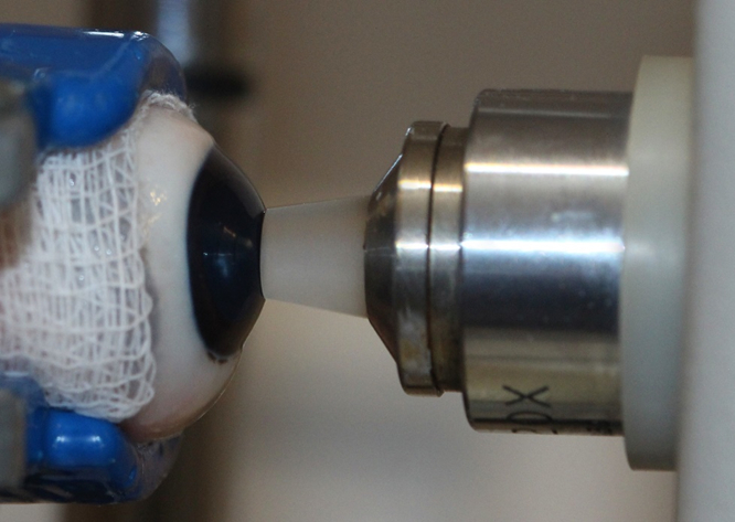
The objective of this study was to determine the endothelial cell density (ECD) and hexagonality of the cornea in the different regions of healthy swine corneal endothelium using specular microscopy. Twenty-four eyeballs from 12 male, 6-month-old Large White pigs (Sus scrofa domesticus) were studied. Contact specular microscopy was performed in the central, superior, inferior, lateral and medial regions. The corneal parameters analysed in this study were ECD and hexagonality. The ECD in the central region was 1865 cells/mm²; in the upper region, it was 1877 cells/mm², in the lower region, it was 1854 cells/mm², in the lateral region, it was 1847 cells/mm², in the medial region, it was 1831 cells/mm². Hexagonality in the central region, was 53%; in the upper region, it was 54%, in the lower region, it was 54%, in the lateral region, it was 54%, in the medial region, it was 54%. There was no significant difference regarding to the evaluated parameters in all corneal regions evaluated. No statistically significantly differences were observed in ECD and hexagonality between the left and the right eyes. This study demonstrates that ECD and hexagonality of the central cornea area represent the entire endothelial mosaic.
Keywords: cornea; endothelium; morphology; cell count, swine.
Downloads
References
Tuft SJ, Coster DJ. The corneal endothelium. Eye. 1990;4(3):389-424. Available from: doi:10.1038/eye.1990.53.
Saad HA, Terry MA, Shamie N, Chen ES, Friend DF, Holiman JD et al. An easy and inexpensive method for quantitative analysis of endothelial damage by using vital dye staining and adobe photoshop software. Cornea. 2008;27(7):818-824. Available from: doi:10.1097/ico.0b013e3181705ca2.
Smeringaiova I, Merjava SR, Stranak Z, Studeny P, Bednar J, Jirsova K. Endothelial wound repair of the organ-cultured porcine corneas. Current Eye Research. 2018;43(7):856-865. Available from: doi:10.1080/02713683.2018.1458883.
Pigatto JAT, Andrade MC, Laus JL, Santos JM, Brooks DE, Guedes PM, et al. Morphometric analysis of the corneal endothelium of Yacare caiman (Caiman yacare) using scanning electron microscopy. Veterinary Ophthalmology. 2004;7(3):205-208. Available from: doi:10.1111/j.1463-5224.2004.04025.
Selig B, Vermeer KA, Rieger B, Hillenaar T, Hendriks CLL. Fully automatic evaluation of the corneal endothelium from in vivo confocal microscopy. BMC Medical Imaging. 2015;15(1)1-15. Available from: doi:10.1186/s12880-015-0054-3.
McCarey BE, Edelhauser HF, Lynn MJ. Review of corneal endothelial specular microscopy for FDA clinical trials of refractive procedures, surgical devices, and new intraocular drugs and solutions. Cornea. 2008;27(1):1-16. Available from: doi:10.1097/ico.0b013e31815892da.
Van Schaick W, van Dooren BT, Mulder PG, Völker-Dieben HJ. Validity of endothelial cell analysis methods and recommendations for calibration in Topcon SP-2000P specular microscopy. Cornea. 2005;24(5):538-544. Available from: doi:10.1097/01.ico.0000151505.03824.6c.
Gwin RM, Lerner I, Warren JK, Gum G. Decrease in canine corneal endothelial cell density and increase in corneal thickness as functions of age. Investigative Ophthalmology & Visual Science. 1982;22(2):267-271. Available from: https://pubmed.ncbi.nlm.nih.gov/7056641/.
Pigatto JAT, Abib FC, Pereira GT, Barros PSM, Freire CD, Laus JL. Density of corneal endothelial cells in eyes of dogs using specular microscopy. Brazilian Journal of Veterinary Research and Animal Science. 2006; 43(4): 476-480. Available from: doi: 10.11606/issn.1678-4456.bjvras.2006.26462.
Pigatto JAT, Cerva C, Freire CD, Abib FC, Bellini LP, Barros PSM, et al. Morphological analysis of the corneal endothelium in eyes of dogs using specular microscopy. Pesquisa Veterinária Brasileira. 2008;28(9):427-430. Available from: doi:10.1590/s0100-736x2008000900006.
Chan-Ling T, Curmi J. Changes in corneal endothelial morphology in cats as a function of age. Current Eye Research. 1988;7(4):387-392. Available from: doi:10.3109/02713688809031788.
Franzen AA, Pigatto JAT, Abib FC, Albuquerque L, Laus JL. Use of specular microscopy to determine corneal endothelial cell morphology and morphometry in enucleated cat eyes. Veterinary Ophthalmology. 2010;13(4):222-226. Available from: doi:10.1111/j.1463-5224.2010.00787.x.
Coyo N, Peña MT, Costa D, Ríos J, Lacerda R, Leiva M. Effects of age and breed on corneal thickness, density, and morphology of corneal endothelial cells in enucleated sheep eyes. Veterinary Ophthalmology. 2016;19(5):367-372. Available from: doi:10.1111/vop.1230.
Andrew SE, Ramsey DT, Hauptman JG, Brooks DE. Density of corneal endothelial cells and corneal thickness in eyes of euthanatized horses. American Journal of Veterinary Research. 2001;62(4):479-482. Available from: doi:10.2460/ajvr.2001.62.479.
Andrew SE, Willis AM, Anderson DE. Density of corneal endothelial cells, corneal thickness, and corneal diameters in normal eyes of llamas and alpacas. American Journal of Veterinary Research. 2002;63(3):326-329. Available from: doi:10.2460/ajvr.2002.63.326.
Albuquerque L, Pigatto JAT, Pacicco LVR. Analysis of the corneal endothelium in eyes of chickens using contact specular microscopy. Semina: Ciências Agrárias. 2015; 36(2): 4199-4206. Available from: doi:10.5433/1679-0359.2015v36n6supl2p4199.
Bercht BS, Albuquerque L, Araujo ACP, Pigatto JAT. Specular microscopy to determine corneal endotelial cell mosphology and morphometry in chinchillas (Chinchilla lanigera) in vivo. Veterinary Ophthalmology. 2015; 18(1): 137-142. Available from: doi:10.1111/vop.12236.
Morita H. Specular microscopy of corneal endothelial cells in rabbits. Journal of Veterinary Medical Science. 1995;57(2):273-277. Available from: doi:10.1292/jvms.57.273.
Brambatti G, Albuquerque L, Pigatto JAT, Vargas EDB, Neumann CF. Corneal endothelial cell density and morphology in rabbits’ eyes using contact specular microscopy. Ciência Rural. 2017;47(12):1-5. Available from: doi:10.1590/0103-8478cr20170027.
Coyo N, Leiva M, Costa D, Rios J, Peña T. Corneal thickness, endothelial cell density, and morphological and morphometric features of corneal endothelial cells in goats. American Journal of Veterinary Research. 2018;79(10):1087-1092. Available from: doi:10.2460/ajvr.79.10.1087.
Hashimoto C, Kurosaka D, Uetsuki Y. Teaching continuous curvilinear capsulorhexis using a postmortem pig eye with simulated cataract. Journal of Cataract & Refractive Surgery. 2001;27(6):814-816. Available from: doi:10.1016/s0886-3350(00)00728-8.
Kermani O, Oberheide U. Comparative micromorphologic in vitro porcine study of IntraLase and Femto LDV femtosecond lasers. Journal of Cataract & Refractive Surgery. 2008;34(8):1393-1399. Available from: doi:10.1016/j.jcrs.2008.04.037.
Nicholls S, Bailey M, Mitchard L, Dick A. Can the corneal endothelium of the pig proliferate in vivo? Acta Ophthalmologica. 2009;87:0. Available from: doi:10.1111/j.1755-3768.2009.2271.x.
Heichel J, Wilhelm F, Kunert K, Schlueter R, Stuhltraeger U, Hammer T. Influence of microkeratome parameters on the stromal bed and flap edge quality in laser in situ keratomileusis. Clinical Ophthalmology. 2014;8: 61-69. Available from: doi:10.2147/opth.s51200.
Heichel J, Blum M, Duncker GIW, Sietmann R, Kunert KS. Surface quality of porcine corneal lenticules after Femtosecond Lenticule Extraction. Ophthalmic Research. 2011;46(2):107-112. Available from: doi:10.1159/000323814.
Gros-Otero J, Ketabi S, Cañones-Zafra R, Garcia-Gonzalez M, Parafita-Fernandez A, Villa-Collar C, et al. Analysis of corneal stromal roughness after iFS 150 kHz and LenSx femtosecond LASIK flap creation in porcine eyes. Graefe's Archive for Clinical and Experimental Ophthalmology. 2019;257(12):2665-2670. Available from: doi:10.1007/s00417-019-04497-7.
Hara H, Cooper DK. Xenotransplantation - the future of corneal transplantation? Cornea. 2011;30(4):371-378. Available from: doi:10.1097/ICO.0b013e3181f237ef.
Lee SE, Mehra R, Fujita M, Roh DS, Long C, Lee W, et al. Characterization of porcine corneal endothelium for xenotransplantation. Seminars in Ophthalmology. 2014;29(3):127-135. Available from: doi:10.3109/08820538.2013.787104.
Bahn CF, Glassman RM, MacCallum DK, Lillie JH, Meyer RF, Robinson BJ, et al. Postnatal development of corneal endothelium. Investigative Ophthalmology & Visual Science. 1986;27(1):44-51. Available from: https://pubmed.ncbi.nlm.nih.gov/3941037/.
Tamayo-Arango LJ, Baraldi-Artoni SM, Laus JL, Vicenti FAM, Pigatto JAT, Abib FC. Ultrastructural morphology and morphometry of the normal corneal endothelium of adult crossbred pig. Ciência Rural. 2009;39(1):117-122. Available from: doi:10.1590/s0103-84782009000100018.
Clerot LL, Hünning PS, Bettio M, Torikachvili M, Petersen MB, Silva AF, et al. Morphology of endothelial cells from different regions of the swine cornea. Acta Scientiae Veterinariae. 2019; 47:1-6. Available from: doi.org/10.22456/1679-9216.89436.
Zeng Y, Yang J, Huang K, Lee Z, Lee X. A comparison of biomechanical properties between human and porcine cornea. Journal of Biomechanics. 2001;34(4):533-537. Available from:10.1016/s0021-9290(00)00219-0.
Fujita M, Mehra R, Lee SE, Roh DS, Long C, Funderburgh JL, et al. Comparison of proliferative capacity of genetically-engineered pig and human corneal endothelial cells. Ophthalmic Research. 2013;49(3):127-138. Available from: doi:10.1159/000342978.
Collin SP, Collin HB. A comparative study of the corneal endothelium in vertebrates. Clinical and Experimental Optometry. 1998;81(6):245-254. Available from: doi:10.1111/j.1444-0938.1998.tb06744.x.
Stapleton S, Peiffer RL Jr. Specular microscopic observations of the clinically normal canine corneal endothelium. American Journal of Veterinary Research. 1979;40(12):1803-1804. Available from: https://pubmed.ncbi.nlm.nih.gov/525905/.
Price NC, Cheng H. Contact and noncontact specular microscopy. British Journal of Ophthalmology. 1981;65(8):568-574. Available from: doi:10.1136/bjo.65.8.568.
Abib FC, Holzchuh R, Schaefer A, Schaefer T, Godois R. The endothelial sample size analysis in corneal specular microscopy clinical examinations. Cornea. 2012;31(5):546-550. Available from: doi: 10.1097/ICO.0b013e3181cc7961.
Chaurasia S, Vanathi M. Specular microscopy in clinical practice. Indian Journal of Ophthalmology. 2021;69(3):517-524. Available from: doi:10.4103/ijo.IJO_574_20.
Doughty MJ. Subjective vs. objective analysis of the corneal endothelial cells in the rabbit cornea by scanning electron microscopy - a comparison of two different methods of corneal fixation. Veterinary Ophthalmology. 2006;9(2):127-135. Available from: doi:10.1111/j.1463-5224.2006.00449.x.
Huang J, Maram J, Tepelus TC, Modak C, Marion K, Sadda SR, et al. Comparison of manual & automated analysis methods for corneal endothelial cell density measurements by specular microscopy. Journal of Optometry. 2018;11(3):182-191. Available from: doi:10.1016/j.optom.2017.06.001.
Coyo N, Leiva M, Costa D, Molina R, Nicolás O, Ríos J, et al. Endothelial cell density and characterization of corneal endothelial cells in the Tawny Owl (Strix aluco) using specular microscopy. Veterinary Ophthalmology. 2019;22(2):177-182. Available from: doi:10.1111/vop.12578.
Abib FC, Barreto Junior J. Behavior of corneal endothelial density over a lifetime. Journal of Cataract & Refractive Surgery. 2001;27(10):1574-1578. Available from: doi:10.1016/s0886-335.
Amann J, Holley GP, Lee SB, Edelhauser HF. Increased endothelial cell density in the paracentral and peripheral regions of the human cornea. American Journal of Ophthalmology. 2003;135(5):584-590. Available from: doi:10.1016/s0002-9394(02)02237-7.
Müller A, Craig JP, Grupcheva CN, McGhee CN. The effects of corneal parameters on the assessment of endothelial cell density in the elderly eye. British Journal of Ophthalmology. 2004;88(3):325-330. Available from: doi:10.1136/bjo.2003.019315.
Published
How to Cite
Issue
Section
License
Copyright (c) 2023 Brazilian Animal Science/ Ciência Animal Brasileira

This work is licensed under a Creative Commons Attribution 4.0 International License.
Authors who publish with this journal agree to the following terms:
- Authors retain copyright and grant the journal right of first publication with the work simultaneously licensed under a Creative Commons Attribution License that allows others to share the work with an acknowledgement of the work's authorship and initial publication in this journal.
- Authors are able to enter into separate, additional contractual arrangements for the non-exclusive distribution of the journal's published version of the work (e.g., post it to an institutional repository or publish it in a book), with an acknowledgement of its initial publication in this journal.
- Authors are permitted and encouraged to post their work online (e.g. in institutional repositories or on their website) prior to and during the submission process, as it can lead to productive exchanges, as well as earlier and greater citation of published work (See The Effect of Open Access).


