
Use of polymethylmethacrylate plaque as a treatment of cranioschisis associated with meningocele in a Girolando heifer: A case report
DOI:
https://doi.org/10.1590/1809-6891v24e-74519EAbstract
Abstract
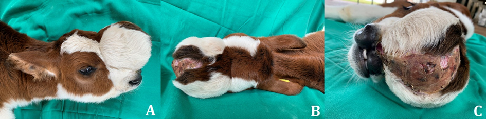
Most genetic diseases affect purebred animals and are inherited as recessive genes. Cranioschisis refers to dysraphism, which occurs in the midline of the skull due to failure to close the cranial symphysis, which can lead to herniation of the meninges filled with cerebrospinal fluid (meningocele), where there is usually a projection of the meningeal tissue. Diagnosis is performed based on clinical examination, characteristic anatomopathological data, and complementary imaging tests. The surgical approach for correction of cranioschisis is the only described as a therapeutic solution and is indicated in cases in which the cranial synthesis defect does not allow for brain protrusion and there is only the occurrence of meningocele, in addition to the absence of severe signs of neurological alteration. This paper reports a case of the use of polymethylmethacrylate (PMMA) plaque to treat cranioschisis associated with meningocele in a Girolando heifer. The surgical opening of the frontonasal sacculation allowed draining a total liquid content of 488 mL, inspection, and suture of the envelope membrane. A PMMA plaque, molded to the bone surface and anchored in the adjacent soft tissue, was used to cover the evidenced frontonasal bone opening. Despite the unfavorable prognosis of the disease, the cranioplasty surgery for the treatment of cranioschisis associated with meningocele using PMMA plaque obtained satisfactory results relative to the quality and maintenance of this animal’s life, evaluated at 19 months postoperatively.
Keywords: bone cement; birth defect; malformation; ruminants.
Downloads
References
Macêdo J, Lucena R, Giaretta P, Kommers G, Fighera R, Irigoyen L, et al. Defeitos congênitos em bovinos da região central do Rio Grande do sul. Pesqui Vet Bras. 2011;31(4):297–306.
Yaman T, Erdogan S, Terzi F, Özyildiz Z. Congenital meningoencephalocele in a Brown Swiss calf : A case report. Eurasian J Vet Sci. 2013;29(2):110–3.
Nogueira D, Bastos R, Rocha L, Rocha E, Nóbrega Neto P, Miranda Neto E, et al. Correção cirúrgica como tratamento de meningocele associada à craniosquise em bezerro: relato de caso. Med Veterinária. 2018;11(4):222.
Zachary J. Sistema Nervoso. In: Zachary, JF; Mcgavin, MD Bases da patologia em veterinária [Internet]. 5th ed. Rio de Janeiro: Elsevier; 2013. p. 774–873. Available from: https://jitc.biomedcentral.com/articles/10.1186/s40425-017-0288-4%0Ahttps://doi.org/10.1007/978-3-0348-9014-4_4%0Ahttps://doi.org/10.1080/00087114.1996.10797378%0Ahttp://www.ncbi.nlm.nih.gov/pubmed/27156851%0Ahttp://dx.doi.org/10.1038/sj.hdy.6800867%0Ahtt
Oliveira D, Medeiros J, Araújo A, Pimentel L, Pierezan F, Neto E, et al. Pulmonary choristoma associated with calf meningocele. Cienc Rural. 2009;39(9):2652–4.
Boscarato A, Pacheco F, Andrade C, Jardim G, Oliveira J, Ribeiro R, et al. Craniosquise with Meningocelis in Newborn Calf. Acta Sci Vet. 2020;48(November):3–8.
Oliveira-Filho J, Badial P, Oliveira A, Álvarez L, Costa J. Cranioplasty for repair of cranioschisis associated with meningocele in a jersey calf. Veterinária e Zootec. 2014;21(3):392–8.
Wronski J, Argenta F, Kemper R, Raiter J, Oliveira N, Driemeier D, et al. Pulmonary choristoma in a new-born calf with multiple cranial and nervous malformations. Cienc Rural. 2022;52(7):1–7.
Back W, Van Den Belt A, Lagerweij E, Van Overbeeke J, Van Der Velden M. Surgical repair of a cranial meningocele in a calf. Vet Rec. 1991;128(24):569–71.
Kohli R, Naddaf H. Surgical treatment of cranial meningocele in Iranian calves. Vet Rec. 1998;142(6):145.
Slimani M, Baus A, Bich CS, de Rousiers A, Duhoux A, Brachet M, et al. Methylmetacrylate (PMMA) cranioplasty technique: Technical interest of intraoperative modeling and review of the literature. Ann Chir Plast Esthet [Internet]. 2022;(xxxx). Available from: https://doi.org/10.1016/j.anplas.2022.09.002
Sanus GZ, Tanriverdi T, Kacira T, Jackson IT. Effects of rigid fixation on the growing neurocranium of immature rabbits. J Craniofac Surg. 2007;18(2):315–24.
Yaremchuk MJ, Fiala TGS, Barker F, Ragland R. The effects of rigid fixation on craniofacial growth of rhesus monkeys. Vol. 93, Plastic and Reconstructive Surgery. 1994. p. 1–10.
Resnick JI, kinney BM, kawamoto JR HK. The Effect of Rigid Internal Fixation on Cranial Growth. Ann Plast Surg. 1990;25(5):372–4.
Southard TE, Franciscus RG, Fridrich KL, Nieves MA, Keller JC, Holton NE, et al. Restricting facial bone growth with skeletal fixation: A preliminary study. Am J Orthod Dentofac Orthop. 2006;130(2):218–23.
Pereira C, Jardim E, Carvalho A, Gealh W, Marão H, Esper H, et al. Técnica cirúrgica para obtenção de enxertos ósseos autógenos intrabucais em reconstruções maxilomandibulares. Rev Bras Cir Craniomaxilofac. 2012;15(2):83–92.
Bayat M, Momen-Heravi F, Khalilzadeh O, Mirhosseni Z, Sadeghi-Tari A. Comparison of conchal cartilage graft with nasal septal cartilage graft for reconstruction of orbital floor blowout fractures. Br J Oral Maxillofac Surg. 2010;48(8):617–20.
Silva T, Araújo-Filho M, Brito M, Freitas R. Mecanismo De Ação, Efeitos Farmacológicos E Reações Adversas Da Ceftriaxona: Uma Revisão De Literatura. Rev Eletrônica Farmácia. 2014;11(3):10.
Machado C, Borges B. Meningite Bacteriana na Unidade de Terapia Intensiva : um Protocolo de Cuidados de Enfermagem. Uniciências. 2015;19(1):79–85.
Ryou M, Coen D. Farmacologia das infecções bacterianas: replicação, transcrição e tradução do DNA. In: Golan DE, Tasgjian-Junior AH, Armstrong EJ, Armstrong AW Princípios de farmacologia - A base fisiopatológica da farmacolterapia. 2nd ed. São Paulo: Guanabara Koogan LTDA; 2014. p. 1215–49.
Published
How to Cite
Issue
Section
License
Copyright (c) 2023 Brazilian Animal Science/ Ciência Animal Brasileira

This work is licensed under a Creative Commons Attribution 4.0 International License.
Authors who publish with this journal agree to the following terms:
- Authors retain copyright and grant the journal right of first publication with the work simultaneously licensed under a Creative Commons Attribution License that allows others to share the work with an acknowledgement of the work's authorship and initial publication in this journal.
- Authors are able to enter into separate, additional contractual arrangements for the non-exclusive distribution of the journal's published version of the work (e.g., post it to an institutional repository or publish it in a book), with an acknowledgement of its initial publication in this journal.
- Authors are permitted and encouraged to post their work online (e.g. in institutional repositories or on their website) prior to and during the submission process, as it can lead to productive exchanges, as well as earlier and greater citation of published work (See The Effect of Open Access).


