
The macroscopic and microscopic description of ruminal lesions in feedlot bovine
DOI:
https://doi.org/10.1590/1809-6891v23e-73109EAbstract
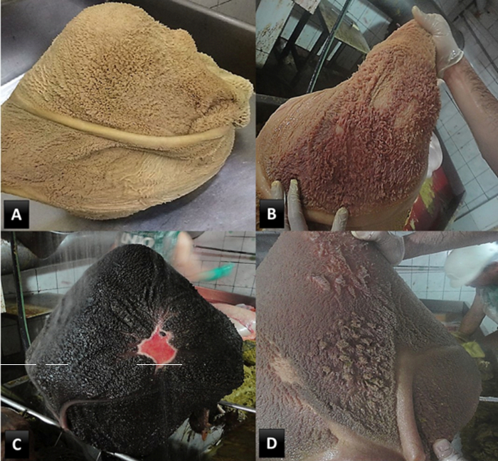
Administration of diets rich in highly fermentable carbohydrates and low fiber content can cause an imbalance between the microorganisms in the rumen with consequent ruminal acidosis. This problem can cause lesions in the rumen wall, often progressing to rumenitis. The purpose of the present was to characterize macroscopic and microscopic ruminal lesions observed in confined feedlot cattle with claw lesions or liver abscess. A total of 1060 bovines were evaluated via post mortem examination. Claw lesions were identified in 88, liver abscess in 10, and macroscopic rumen lesions in 230 bovines; furthermore, 178 rumens were characterized with hyperkeratosis, 41 with hyperemia, 9 with ulcer, and 2 with neoplasia. The 98 bovines with claw lesions and liver abscess were selected for histopathological examination. Of these, macroscopic lesions were noted in 23 and microscopic lesions in 23 animals. Of the 23 animals that presented macroscopic lesions, 10 showed the same changes observed under microscopy. Seven cases of hyperkeratosis were diagnosed in the macro and microscopic evaluation. Of the 5 cases of hyperemia verified on macroscopy, 2 cases were identified via microscopy, and 1 case of ulcer identified through macroscopy and microscopy. The microscopic evaluation of the rumens allowed the identification of lesions in animals with claw lesions that did not present macroscopic rumen alterations.
Keywords: cattle; fridge; histopathology; rumen; rumenitis.
Downloads
References
Neto JAS, Oliveira VS, Santos ACP, Valença RL. Distúrbios metabólicos em ruminantes- uma revisão. Ver. Bras. Hig. San. Anim. 2014; 8(4): 157-86.
Vechiato TAF, Maschio W, Bom LC, Lopes PD, Ortolani EL. Estudo retrospectivo de abscessos hepáticos em bovinos abatidos em um frigorífico paulista. Bras. J. Vet. Anim. Sci. 2011; 48(5): 384-91.
Nagaraja TG, Lechtenberg KF. Liver abscesses in feedlot cattle. Vet. Clin. Food Anim. 2007; 23: 351-69.
Plaizier JC, Khafipour E, Li S, Gozho GN, Krause DO. Subacute ruminal acidosis (SARA), endotoxins and health consequences. Anim. Feed Sci. Technol. 2012; 172: 9-21.
Wang DS, Zhang RY, Zhu WY, Mao SY. Effects of subacute ruminal acidosis challenges on fermentation and biogenic amines in the rumen of dairy cows. Livest. Sci. 2013; 155: 262-72.
Radostits OM, Gay CC, Hinchcliff KW, Constable PD. Veterinary medicine. 3 ed. St. Louis: Elsevier, 2007, 2156p.
Tadepalli S, Narayanan SK, Stewart GC, Chengappa MM, Nagaraja TG. Fusobacterium necrophorum: A ruminal bacterium that invades liver to cause abscesses in cattle. Anaerobe. 2009; 15: 36-43.
Narayanan S, Nagaraja TG, Okwumabua OGI, Staats J, Chengappa MM, Oberst RD. Ribotyping to compare Fusobacterium necrophorum isolates from bovine liver abscesses, ruminal walls, and ruminal contents. Appl. Environ. Microbiol. 1997; 63(12): 4671-78.
Nocek, JE. Bovine acidosis: implications on laminitis. J Dairy Sci. 1997; 80: 1005-28.
Owens FN, Secrist DS, Hill WJ, Gill DR. Acidosis in cattle: a review. J. Anim. Sci. 1998; 76: 275-86.
Vechiato, TAF, Maschio, W, Bom, LC, Lopes, PD, Ortolani, EL. Retrospective study of liver abscesses in beef cattle slaughtered in a Brazilian abattoir. Brazilian Journal of Veterinary Research and Animal Science, 2011; 48(5): 384-391.
Habel RE. Sistema digestivo do ruminante. In Getty R, Grossman S. Anatomia dos animais domésticos. Rio de Janeiro: Guanabara Koogan, 1986, 858p.
Samuelson DA. Tratado de histologia veterinária. Rio de Janeiro: Elsevier, 2007, 544p.
Brasil. Decreto n° 30.691 de 29 mar 1952. Aprova o novo regulamento da inspeção industrial e sanitária de produtos de origem animal. Ministério da Agricultura, Pecuária e Abastecimento. Diário oficial da União, Brasília (7 jul 1952).
Tolosa EMC, Rodrigues CJ, Behmer OA, Freitas Neto AG. Manual de técnicas para histologia normal e patológica. Barueri: Manole, 2003, 331p.
Sampaio IBM. Estatística aplicada à experimentação animal. Belo Horizonte: FEP-MZV, 2007, 265p.
Nagaraja, TG, Lechtenberg KF. Acidosis in feedlot catlle. Vet. Clin. Food Anim. 2007; 23: 333-50.
Nagaraja TG. Rumen health. In Simpósio de nutrição de ruminantes – saúde do rúmen. Anais eletrônicos [CD-ROM]. Botucatu: Unesp, 2011.
Hernández J, Benedito JL, Abuelo A, Castillo C. Ruminal acidosis in feedlot: From aetiology to prevention. Scientific World J. 2014; article ID 702572 [acesso 01 mai 2017]. Disponível em: http://dx.doi.org/10.115.5/2014/702572
Steele MA, Croom J, Kahler M, AlZahal O, Hook SE, Plaizier K, McBride BW. Bovine rumen epithelium undergoes rapid structural adaptations during grain-induced subacute ruminal acidosis. Am. J. Physiol. Regul. Integr. Comp. Physiol. 2011; 300: 1515-23.
Tessele B, Barros CSL. Tumores em bovinos encontrados em abatedouros frigoríficos. Pesq. Vet. Bras. 2016; 36(3): 145-60.
Bertone AL. Neoplasms of the bovine gastrointestinal tract. Vet. Clin. North Am. Food Anim. Pract. 1990; 6 (2): 515-24.
Correia WM, Correia CNM. Doenças infecciosas em animais domésticos. São Paulo: Verela, 1992, 843p.
Valente TNP, Sampaio CB, Lima ES, Deminicis BB, Cezário AS, Santos WBR. Aspects of acidosis in ruminants with a focus on nutrition: a review. J. Agr. Sci. 2017; 9(3): 90-97.
Rezac DJ, Thomson DU, Bartle SJ, Osterstock JB, Prouty FL, Reinhardt CD. Prevalence, severity, and relationships of lung lesions, liver abnormalities, and rumen health scores measured at slaughter in beef cattle. J. Anim. Sci. 2014; 92: 2595-602.
Smith HA. Ulcerative lesions of the bovine rumen and their possible relation to hepatic abscess. Am. J. Vet. Res. 1944; 5: 234-42.
Costa SF, Pereira MN, Melo LQ, Caliari MV, Chaves ML. Alterações morfológicas induzidas por butirato, propionato e lactato sobre a mucosa ruminal e epiderme de bezerros. II. Aspectos ultra-estruturais. Arq. Bras. Med. Vet. Zootec. 2008; 60(1): 10-18.
Xu Y, Ding Z. Physiological, biochemical and histopathological effects of fermentative acidosis in ruminant production: a minimal review. Span J Agric Res. 2011; 9(2): 414-22.
González LA, Manteca X, Calsamiglia S, Schwartzkopf GKS, Ferret A. Ruminal acidosis in feedlot cattle: Interplay between feed ingredients, rumen function and feeding behavior. Anim. Feed Sci. Technol. 2012; 172: 66-79.
Borges, JRJ. et al. Doenças dos dígitos dos bovinos: nomenclatura padronizada para o Brasil. Revista CFMV, Brasília, v.23, n.73, p.45-72, abr./jun. 2017.
Egger-Danner et al. ICAR claw health atlas. https://www.icar.org/ICAR_Claw_Health_Atlas.pdf, 2020.
Published
How to Cite
Issue
Section
License
Copyright (c) 2022 Brazilian Animal Science/ Ciência Animal Brasileira

This work is licensed under a Creative Commons Attribution 4.0 International License.
Authors who publish with this journal agree to the following terms:
- Authors retain copyright and grant the journal right of first publication with the work simultaneously licensed under a Creative Commons Attribution License that allows others to share the work with an acknowledgement of the work's authorship and initial publication in this journal.
- Authors are able to enter into separate, additional contractual arrangements for the non-exclusive distribution of the journal's published version of the work (e.g., post it to an institutional repository or publish it in a book), with an acknowledgement of its initial publication in this journal.
- Authors are permitted and encouraged to post their work online (e.g. in institutional repositories or on their website) prior to and during the submission process, as it can lead to productive exchanges, as well as earlier and greater citation of published work (See The Effect of Open Access).


