
Histological and immunohistochemical evaluations of the bone marrow from femur and sternal manubrium of dogs reactive for leishmaniasis by DPP® and ELISA tests
DOI:
https://doi.org/10.1590/1809-6891v23e-73104EAbstract
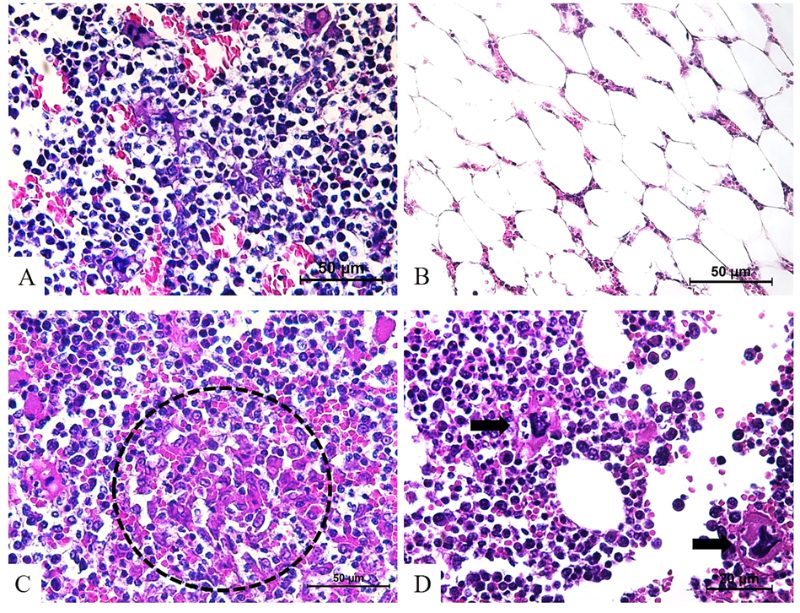
As the bone marrow is one of the most organs affected by canine visceral leishmaniasis (CVL), samples from this are frequently taken for parasitological tests, with occurrence of myelodysplastic changes, with consequent anemia, leukopenia, and thrombocytopenia. Thus, this study aimed to investigate the histological and immunohistochemical changes in the bone marrow of the femur and sternal manubrium of dogs reactive for leishmaniasis by DPP® and ELISA tests. For this, thirteen canines from the epidemiological routine for CVL carried out by the Directorate of Zoonosis Surveillance of Goiânia (DVZ), GO, Brazil, were subjected to anatomopathological examination. 46.2% of bone marrow samples from the femur showed a higher proportion of the red series, and 53.9% of bone marrow of the sternal manubrium evidenced a higher proportion of the red series. Also, there were varied macrophage hyperplasia, hemosiderosis, and megakaryocytic emperipolesis. Amastigote forms of Leishmania spp. in the bone marrow of the femur and sternal manubrium to histopathological and immunohistochemical evaluations were observed, with good agreement them, but without difference in the parasite intensity between the bone marrow of these anatomical sites. It was concluded that bone marrow of the femur and sternal manubrium of dogs reactive for leishmaniasis by DPP® and ELISA tests has histological changes resulting from the disease, regardless of the parasite presence or intensity, with macrophage hyperplasia, hemosiderosis, and emperipolesis being the main medullary changes in these animals. Also, the bone marrow of the femur and sternal manubrium are useful anatomical sites for the diagnosis of CVL by direct methods.
Keywords: amastigotes; canine visceral leishmaniasis; histopathology; immunostaining; medullary changes
Downloads
References
World Health Organization. Control of the leishmaniases; 2016. 375p.
Ministério da Saúde. Leishmaniose visceral: o que é, causas, sintomas, tratamento, diagnóstico e prevenção. Situação epidemiológica da leishmaniose visceral. 2020. [cited 2020 jul 20]. Available from: http://www.saude.gov.br/saude-de-a-z/leishmaniose-visceral. Portuguese.
Coura-Vital W, Marques MJ, Veloso VM, Roatt BM, Aguiar-Soares RDO, Reis LES, et al. Prevalence and factors associated with Leishmania infantum infection of dogs from an urban area of Brazil as identified by molecular methods. Plos Neglected Tropical Diseases. [Internet]. 2011 aug [cited 2020 jun 25], 5(8):e1291. Available from: https://doi.org/10.1371/journal.pntd.0001291
Castro-Júnior JG, Freire ML, Campos SPS, Scopel KKG, Porrozzi R, Silva ED, et al. Evidence of Leishmania (Leishmania) infantum infection in dogs from Juiz de Fora, Minas Gerais State, Brazil, based on immunochromatographic dual-path platform (DPP®) and PCR assays. Revista do Instituto de Medicina Tropical de São Paulo. [Internet]. 2014 may [cited 2020 jun 28]; 56(3): 225-229. Available from: doi: https://doi.org/10.1590/S0036-46652014000300008
Ministério da Saúde. Secretaria de Vigilância em Saúde. Departamento de Vigilância Epidemiológica. Manual de vigilância e controle da leishmaniose visceral; 2014.[cited 2020 jul 20];120 p. Available from: https://bvsms.saude.gov.br/bvs/publicacoes/manual_vigilancia_controle_leishmaniose_visceral_1edicao.pdf. Portuguese
Menezes RC, Madeira MF, Ferreira LC, Barbosa CJLF, Miranda LHM, Figueiredo FB. Cell-block immunohistochemistry of bone marrow aspirates: a novel tool to improve the diagnosis of Leishmania infection in dogs. Journal of comparative pathology. [Internet]. 2016. Feb-Apr [cited 2020 jul 28]; 154(2-3):157-60. Available from; doi: 10.1016/j.jcpa.2015.12.005
Paparcone R, Fiorentino E, Cappiello S, Gizzarelli M, Gradoni L, Oliva G, Manzillo VF. Sternal aspiration of bone marrow in dogs: a practical approach for canine leishmaniasis diagnosis and monitoring. Journal of Veterinary Medicine. [Internet]. 2013.[cited 2020 jul 29]; 2013: 217314. Available from: https://doi.org/10.1155/2013/217314
Tafuri WL, Santos RL, Arantes RME, Gonçalves R, Melo MN, Michalik MSM, et al An alternative immunohistochemical method for detecting Leishmania amastigotes in paraffin-embedded canine tissues. Journal of immunological methods. [Internet]. 2004, Sep [cited 2020 jul 15]; 292(1-2):17-23. Available from: doi: 10.1016/j.jim.2004.05.009
Keenan CM, Hendricks LD, Lightner L, Johnson AJ. Visceral leishmaniasis in the German Shepherd dog. II. Pathology. Veterinary pathology. [Internet]. 1984 Jan [cited 2020 jul 19]; 21(1):80-6. Available from: doi: 10.1177/030098588402100114
Lima WG, Michalick MSM, Melo MN, Tafuri WL, Tafuri WL. Canine visceral leishmaniasis: a histopathological study of lymph nodes. Acta Tropica. [Internet]. 2004. Sep [cited 2020 jul 26]; 92(1):43-53. doi: 10.1016/j.actatropica.2004.04.007.
Cavalcanti AS, Ribeiro-Alves M, Pereira LOR, Mestre GL, Ferreira ABR, Morgado FN, et al. Parasite load induces progressive spleen architecture breakage and impairs cytokine mRNA expression in Leishmania infantum-naturally infected dogs. Plos one. [Internet]. 2015, apr [cited 2020 jul 26]; 10(4):e0123009. Available from: doi: 10.1371/journal.pone.0123009
Xavier SC, Andrade HM, Monte SJH, Chiarelli IM, LimaWG, Michalick MSM, et al. Comparison of paraffin-embedded skin biopsies from different anatomical regions as sampling methods for detection of Leishmania infection in dogs using histological, immunohistochemical and PCR methods. BMC Veterinary Research. [Internet]. 2006. 2:17. [cited 2020 de jul de 20]; 8;2:17. Availables from: doi: 10.1186/1746-6148-2-17
Tryphonas L, Zawidzka Z, Bernard MA, Janzen EA. Visceral Leishmaniasis in a dog: clinical, hematological and pathological observations. Canadian Journal of Comparative Medicine. [Internet]. 1977 jan [cited 2020 jul 30]; 41(1):1-12. Available from: Disponível em: https://www.ncbi.nlm.nih.gov/pmc/articles/PMC1277686/
Reis AB, Martins OAF, Teixeira-Carvalho A, Giunchetti RC, Carneiro CM, Mayrink W, et al. Systemic and compartmentalized immune response in canine visceral leishmaniasis. Veterinary Immunology and Immunopathology. [Internet]. 2009 mar [cited 2020 jul 28]; 128(1-3):87-95. Available from: https://doi.org/10.1016/j.vetimm.2008.10.307
Mahajan V, Marwaha RK. Case report immune mediated hemolysis in visceral leishmaniasis. Journal of Tropical Pediatrics. [Internet]. 2007 aug [cited 2020 jul 30]; 53(4):284–286. Available from: https://doi.org/10.1093/tropej/fmm018
Momo C, Jacintinho APP, Moreira PRR, Munari DP, Machado GF, Vasconcelos RO. Morphological changes in the bone marrow of the dogs with visceral leishmaniasis. Veterinary Medicine International. [Internet]. 2014 [cited 2020 jul 12] 2014: 5p. Available from: http://dx.doi.org/10.1155/2014/150582
Solimando AG, Coniglio G, Desantis V, Lauletta G, Bavaro DF, Diella L, et al. A Challenging Case of Visceral Leishmaniasis. Reports. [Internet]. 2002 [cited 2020 aug 30]; 5(2):23. Available from: https://doi.org/10.3390/reports5020023
Toplu N, Aydogan A. An immunohistochemical study in cases with usual and unusual clinicopathological findings of canine visceral leishmaniosis. Parasitology research. [Internet]. 2011 oct [cited 2020 jul 29]; 109(4):1051-7. Available from: doi: 10.1007/s00436-011-2345-0
Oliveira VC, Boechat VC, Mendes AAVJ, Madeira MF, Ferreira LC, Figueiredo FB, et al. Occurrence of Leishmania infantum in the central nervous system of naturally infected dogs: Parasite load, viability, co-infections and histological alterations. Plos one. [Internet]. 2017 apr [cited 2020 jul 20]; 12(4):e0175588. Available from: doi: 10.1371/journal.pone.0175588
Castro MC, Vieira AB, Santos MCS, Gershony LC, Soares AMB, Ferreira AMR. Escore de condição corporal como indicador do prognóstico de gatos com doença renal crônica. Ciência Rural [Internet], 2010 fev [cited 2020 jul 20]; 40(2):365-370. Available from: https://doi.org/10.1590/S0103-84782010005000010 .Portuguese
Lima IS, Silva JS, Almeida VA, Leal GLJ, Souza PAN, Larangeira DF, Moura-Neto JP, et al. Severe clinical presentation of visceral leishmaniasis in naturally infected dogs with disruption of the splenic White pulp. Plos One. [Internet]. 2014.fev [cited 2020 jul 28]; 3;9(2):e87742. Available from: doi: 10.1371/journal.pone.0087742
Boechat VC, Mendes AAVJ, Madeira MF, Ferreira LC, Figueiredo FB, Rodrigues FCC, et al. Occurrence of Leishmania infantum and associated histological alterations in the genital tract and mammary glands of naturally infected dogs. Parasitology research. [Internet]. 2016.jun [cited 2020 jul 20]; 115(6):2371-9. Available from: doi: 10.1007/s00436-016-4987-4.
Caputo LFG, Gitirana LB, Manso PPA. Capítulo 3. Técnicas histológicas. IN: Molinaro EM, Caputo LFG, Amendoeira MRR. Conceitos e métodos para a formação de profissionais em laboratórios de saúde: volume 2 / Rio de Janeiro: EPSJV; IOC, 2010, p.89-188. Portuguese. (https://www.epsjv.fiocruz.br/publicacao/livro/conceitos-e-metodos-para-formacao-de-profissionais-em-laboratorios-de-saude-volum-2) . Portuguese
Nunes CS, Cinsa LA. Princípios Do Processamento Histológico De Rotina. Revista Interdisciplinar de Estudos Experimentais, v. 8, n. único, p. 31-40, 2016. Portuguese (https://docs.bvsalud.org/biblioref/2018/11/964830/2884-8890-1-sm.pdf)
Santana CC, Vassallo J, Freitas LAR, Oliveira GGS, Pontes-de-Carvalho LC, Dos-Santos WLC. Inflammation and structural changes of splenic lymphoid tissue in visceral leishmaniasis: A study on naturally infected dogs. Parasite Immunology. [Internet]. 2008 oct [cited 28 jul 2020]; 30(10): 515–524. Available from: doi: 10.1111/j.1365-3024.2008.01051.x
Silva AVA, Figueiredo FB, Menezes RC, Mendes-Junior AA, Miranda LHM, Cupolillo E. Morphophysiological changes in the splenic extracellular matrix of Leishmania infantum-naturally infected dogs is associated with alterations in lymphoid niches and the CD4+T cell frequency in spleens. PLoS neglected tropical diseases [Internet]. 2018 apr [cited 2020 jul 20]; 12(4):e0006445. Available from: https://doi.org/10.1371/journal.pntd.0006445
Tafuri WL, Tafuri WL, Barbosa AJA, Michalick MSM, Genaro O, França-Silva JC. Histopathology and immunocytochemical study of type 3 and type 4 complement receptors in the liver and spleen of dogs naturally and experimentally infected with Leishmania (Leishmania) chagasi. Revista do Instituto de Medicina Tropical de São Paulo [Internet]. 1996 apr [cited 2020 jul 25]; 38(2):81-89. [acesso 25 de julho de 2020]. Available from: http://dx.doi.org/10.1590/S0036-46651996000200001
Reis AB, Martins-Filho AO, Teixeira-Carvalho A, Carvalho MG, Mayrink W, França-Silva JC. Parasite density and impaired biochemical/hematological status are associated with severe clinical aspects of canine visceral leishmaniasis Research in Veterinary Science. [Internet]. 2006 aug [cited 2020 jul 25]; 81:68-75. Available from: https://doi.org/10.1016/j.rvsc.2005.09.011
Yarali N, Fişgin T, Duru F, Kara A. Myelodysplastic features in visceral leishmaniasis. American Journal of Hematology. [Internet]. 2002 oct [cited 2020 jul 26]; 71:191-195. Available from: doi: 10.1002/ajh.10200
Manzillo VF, Restucci B, Pagano A, Gradoni, Oliva G. Pathological changes in the bone marrow of dogs with leishmaniosis. The Veterinary Record. [Internet]. 2006, may [cited 2020 jul 20]; 158:690-694. Available from: http://dx-doi-org.ez49.periodicos.capes.gov.br/10.1136/vr.158.20.690
Sable MN, Sehgal K, Gadage VS, Subramanian PG, Gujral S. Megakaryocytic emperipolesis: A histological finding in myelodysplastic syndrome. Indian journal of pathology & microbiology. [Internet]. 2009 oct-dec [cited 2020 jul 26]; 52(4):599-600. Available from: 10.4103/0377-4929.56153
Anosa VO, Logan-Henfrey LL, Shaw MK. A light and electron microscopic study of changes in blood and bone marrow in acute hemorrhagic Trypanosoma vivax infection in calves. Veterinary pathology. [Internet] 1992 jan [cited 2020 aug 31]; 29(1):33-45. Available from: doi:10.1177/030098589202900105
Cunin P, Nigrovic PA. Megakaryocyte emperipolesis: a new frontier in cell-in-cell interaction. Platelets.[Internet] 2020 aug [cited 2020 aug 30]; 31(6):700-706. Available from: doi:10.1080/09537104.2019.1693035
Menezes RC, Figueiredo FB, Wise AG, Madeira MF, Oliveira RVC, Schubach TMP, et al. Sensitivity and specificity of in situ hybridization for diagnosis of cutaneous infection by Leishmania infantum in dogs. Journal of Clinical Microbiology. [Internet]. 2013 jane [cited 2020 jul 28]; 51:206-211. Available from: doi: 10.1128/JCM.02123-12
Queiroz NMGP, Silveira RCV, Noronha ACFJ, Oliveira TMFS, Machado RZ, Starke-Buzetti WA. Detection of Leishmania (L.) chagasi in skin. Veterinary Parasitology. [Internet]. 2011 may [cited 2020 jul 28]; 178:1-8. Available from: doi:10.1016/j.vetpar.2010.12.033
Quintella LP, Cuzzi T, Madeira MF, Okamoto T, Schubach AO. Immunoperoxidase technique using an anti-Leishmania (L.) chagasi hyperimmune serum in the diagnosis of culture- confirmed american tegumentary leishmaniasis. Revista do Instituto de Medicina Tropical de São Paulo. [Internet]. 2009 apr [cited 2020 jul 28] 51(2): 83-86. Available from: doi:10. 1590/S0036-46652009000200005
Published
How to Cite
Issue
Section
License
Copyright (c) 2022 Brazilian Animal Science/ Ciência Animal Brasileira

This work is licensed under a Creative Commons Attribution 4.0 International License.
Authors who publish with this journal agree to the following terms:
- Authors retain copyright and grant the journal right of first publication with the work simultaneously licensed under a Creative Commons Attribution License that allows others to share the work with an acknowledgement of the work's authorship and initial publication in this journal.
- Authors are able to enter into separate, additional contractual arrangements for the non-exclusive distribution of the journal's published version of the work (e.g., post it to an institutional repository or publish it in a book), with an acknowledgement of its initial publication in this journal.
- Authors are permitted and encouraged to post their work online (e.g. in institutional repositories or on their website) prior to and during the submission process, as it can lead to productive exchanges, as well as earlier and greater citation of published work (See The Effect of Open Access).


