
Evaluation of mesenchymal cells and dapsone for the treatment of dermonecrotic wounds caused by Loxosceles laeta venom in rabbits
DOI:
https://doi.org/10.1590/1809-6891v23e-72573EAbstract
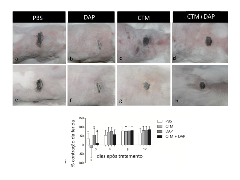
This study aimed to evaluate the efficacy of mesenchymal stem cells (MSC), alone or associated with dapsone (DAP), in treating dermonecrotic wounds caused by Loxosceles laeta venom. Twenty-five male rabbits were distributed into five groups. Negative control received ultrapure water (C-), whilst all other groups were injected with 20 μg of L. laeta venom. After 4 hours, each group received one of the following treatments: PBS (C+), DAP, MSC, and DAP+MSC. Animals were evaluated daily and photographic records made for analysis of wound area. Twelve days after, animals were euthanized and skin samples removed for histological analysis. We observed that DAP showed the best percentage of wound contraction at day 3. In the treatments using MSCs, a negative value of wound contraction was observed for the isolated MSCs, as well as a lower contraction value for the association of the MSC + DAP when compared to PBS, probably, by the increase in initial inflammation after the application of stem cells, due to the fact that MSCs secrete a broad spectrum of bioactive molecules such as cytokines and growth factors that favor regeneration. Histologically, it was observed that animals of C+ showed extensive areas of necrosis, ulcers, neutrophilic infiltrate, and mineralization. Collagen deposition showed increase in MSC+DAP treatment, however vascularization remained unchanged. This is the first report using MSC and MSC+DAP as a treatment for cutaneous loxoscelism and more studies are needed to determine its use as an alternative therapy for dermonecrotic lesions caused by Loxosceles spider.
Key words: loxoscelism; MSC; stem cells; spider venom; wound repair.
Downloads
References
Andrade RMG, Lourenço WR, Tambourgi DV. Comparison of the fertility between Loxosceles intermedia and Loxosceles laeta spiders (Araneae, Sicariidae). 2000. Journal of Arachnology. 2000; 28, 245-247.
Malaque CMS, Castro-Valencia JE, Cardoso JLC, França FOS, Barbaro K, Fan HW. Clinical and epidemiological features of definitive and presumed loxoscelism in São Paulo, Brazil. Revista do Instituto de Medicina Tropical de São Paulo. 2002; 44,139-143. Disponível em: http://dx.doi.org/10.1590/S0036-46652002000300005.
Miranda, ALS, Guerra-Duarte C., Lima, SA, Chávez-Olórtegui C, Soto-Blanco B. History, challenges and perspectives on Loxosceles (brown spiders) antivenom production in Brazil. Toxicon. 2021, 40-45. Disponível em: https://doi.org/10.1016/j.toxicon.2021.01.004
Silva PH, Silveira RB, Appel MH, Mangili OC, Gremisk W, Veiga SS. Brown spiders and loxoscelism. Toxicon. 2004.44, 693-709. https://doi.org/10.1016/j.toxicon.2004.07.012.
Smith CW, Micks DW. The role of polymorphonuclear leukocytes in the lesion caused by the venom of the brown spider. Laboratory Investigation 1970. 22, 90-93.
Tambourgi DV, Gonçalves-de-Andrade RM, van den Berg CW. Loxoscelism: From basic research to the proposal of new therapies. Toxicon. 2010. 15;56(7):1113-9. Disponível em: https://doi.org/10.1016/j.toxicon.2010.01.021.
Malaque CMS, Santoro ML, Cardoso JL, Conde MR, Novaes CTG, Risk JY, França FOS, Medeiros CR, Fan HW. Clinical picture and laboratorial evaluation in human loxoscelism. Toxicon. 2011. 58, 664-671. Disponível em: https://doi.org/ 10.1016/j.toxicon.2011.09.011.
Barbaro KC, Knysak I, Martins R, Hogan C, Winkel K. Enzymatic characterization, antigenic cross-reactivity and neutralization of dermonecrotic activity of five Loxosceles spider venoms of medical importance in the Americas. Toxicon. 2005. 45, 489-499. Disponível em: https://doi.org/10.1016/j.toxicon.2004.12.009.
Ministério da saúde, Brasil. Manual de diagnóstico e tratamento de acidentes por animais peçonhentos. Brasília: Assessoria de comunicação e educação em saúde – Fundação Nacional de Saúde. 2001. 45-56.
Peterson ME. Brown Spider Envenomation. Clinical techniques in Small Animal Practice. 2006. 21, 191-193. Disponível em: https://doi.org/10.1053/j.ctsap.2006.10.004.
Pauli I, Minozzo JC, Silva PH, Chaim OM, Veiga SS. Analysis of therapeutic benefits of antivenin at different time intervals after experimental envenomation in rabbits by venom of the brown spider (Loxosceles intermedia). Toxicon. 2009. 53, 660-671. Disponível em: https://doi.org/10.1016/j.toxicon.2009.01.033.
Costa TGF, Costal-Oliveira F, de Assis TCS, Lima SA, Martins CA, Finco AB, Veiga SS, Soccol VT, Machado-de-Ávila RA, Figueiredo LFM, Minozzo JC, Kalapothakis E, Guerra-Duarte C, Alvarenga LM, Chávez-Olórtegui C. Engineered antigen containing epitopes from Loxosceles spp. spider toxins induces a monoclonal antibody (Lox-mAb3) against astacin-like metalloproteases. International Journal of Biological Macromolecules. 2020. Nov 1;162:490-500. Disponível em: https://doi.org/10.1016/j.ijbiomac.2020.06.176.
Saavedra-Langer R, Costa TGF, Lima SA, Costal-Oliveira F, Martins CA, Machado-de-Ávila RA, Minozzo JC, Soccol VT, Guerra-Duarte C, Kalapothakis E, Chávez-Olórtegui C. A prokaryote system optimization for rMEPLox expression: A promising non-toxic antigen for Loxosceles antivenom production. International Journal of Biological Macromolecules. 2021 Sep 30;187:66-75. Disponível em: https://doi.org/10.1016/j.ijbiomac.2021.07.042.
Fortier LA. Stem cells: classifications, controversies, and clinical applications. Veterinary Surgery. 2005. 34, 415-423. Disponível em: https://doi.org/10.1111/j.1532-950X.2005.00063.x.
Minguell JJ, Erices A, Conget P. Mesenchymal stem cells. Experimental Biology and Medicine. 2001. 226, 507-520.
Jeong JH. Adipose Stem cells and skin repair. Current Stem Cell Research and Therapy. 2010. 5, 137-140.2010.
Gaur M, Dobke M, Lunyak VV. Mesenchymal stem cells from adipose tissue in clinical applications for dermatological indications and skin aging. International Journal of Molecular Sciences. 2017. 18, 208. Disponível em: https://doi.org/10.3390/ijms18010208.
Kim, W., Park, B., Sung, J., Yang, J., Park, S., Kwak, S., Park, J. Wound healing effect of adipose-derived stem cells: A critical role of secretory factors on human dermal fibroblasts. Journal of Dermatological Science. 2007. 48, 15-24. Disponível em: https://doi.org/10.1016/j.jdermsci.2007.05.018.
Kasperk C, Wergedal J, Strong D, Farley J, Wangerin K, Gropp H, Ziegler R, Baylink DJ. Human bone cell phenotypes differ depending on their skeletal site of origin. The Journal of Clinical Endocrinology and Metabolism. 1995. 80, 2511-2517. Disponível em: https://doi.org/10.1210/jcem.80.8.7629252.
Phillips S, Kohn M, Baker D, Vander Leest R, Gomez H, McKinney P, McGoldrick J, Brent J. Therapy of brown spider envenomation: a controlled trial of hyperbaric oxygen, dapsone, and cyproheptadine. Annals of Emergency Medicine.1995 Mar;25(3):363-8. Disponível em: https://doi.org/10.1016/s0196-0644(95)70296-2. PMID: 7864478.
Oliveira ST, Leme MC, Pippi NL, Raiser AG, Manfron MP. Preparations of comfrey (Symphytum officinale) on cutaneous wound healing in rats. Revista da FZVA. 2000. 7, 65-74
Wu Y, Chen L, Scott PG, Tredget EE. Mesenchymal stem cells enhance wound healing through differentiation and angiogenesis. Stem Cells. 2007. 25, 2648-2659.
Zhang J, Huang X, Wang H, Liu X, Zhang T, Wang Y, Hu D. The challenges and promises of allogeneic mesenchymal stem cells for use as a cell-based therapy. Stem Cell Research and Therapy. 2015. Disponível em: https://doi.org/10.1186/s13287-015-0240-9
Ferrara GIS, Fernandes-Pedrosa MF, Azevedo ILMJ, Andrade RMG, Portaro FCV, Almeida DM, Murakami M, Arni RK, Berg CW, Ho L, Tambourgi DV. Smase II, a new sphingomyelinase D from Loxosceles laeta venom gland: Molecular cloning, expression, function and structural analysis. Toxicon. 2009. 53, 743-753. Disponível em: https://doi.org/10.1016/j.toxicon.2009.02.013.
Elston DM, Miller SD, Young RJ, Eggers J, Mcglasson D, Schmidt WH, Bush A. Comparison of colchicine, dapsone, triamcinolone, and diphenhydramine therapy for the treatment of brown recluse spider envenomation. Archives of Dermatology. 2005. 141, 595-597. Disponível em: https://doi.org/10.1001/archderm.141.5.595.
Caplan AI. Adult mesenchymal stem cells for tissue engineering versus regenerative medicine. Journal of Cellular Physiology. 2007. 213, 341-347. Disponível em: https://doi.org/10.1002/jcp.21200.
Harman RJ. Stem cell therapy in veterinary dermatology. Veterinary Dermatology. 2013. 24, 90-e24. Disponível em: https://doi.org/10.1111/vde.12000.
Kim JW, Lee JH, Lyoo YS, Jung DI, Park HM. The effects of topical mesenchymal stem cell transplantation in canine experimental cutaneous wounds. Veterinary Dermatology. 2013. 24, 242-253. Disponível em: https://doi.org/10.1111/vde.12011.
Ospedal KZ, Appel MH, Neto JF, Mangili OC, Sanches V, Gremski W. Histopathological findings in rabbits after experimental acute exposure to the Loxosceles intermedia (brown spider) venom. International Journal of Experimental Pathology. 2002. 83, 287-294.
Maynor ML, Moon RE, Klitzman B, Fracica PJ, Canada A. Brown recluse spider envenomation: a prospective trial of hyperbaric oxygen therapy. Academic Emergency Medicine. 1997.4, 184-192.
Rees RS, Altenbern DP, Lynch JB, King Jr LE. Brown recluse spider bites. A comparison of early surgical excision versus dapsone and delayed surgical excision. Annals of Surgery. 1985. 202, 659-663.
Booth SA, Moody CE, Dahl MV, Herron MJ, Nelson RD. Dapsone suppresses integrin-mediated neutrophil adherence function. Journal of Investigative Dermatology. 1992. 98, 135-40.
Barret SM, Jenkings MR, Fisher DE. Dapsone or electric Shock therapy of brown recluse spider envenomation? Annals of Emergency Medicine. 1994. 24, 21-25.
Hogan CJ, Barbaro KC, Winkel K. Loxoscelism: old obstacles, new directions. Annals of Emergency Medicine. 2004. 44, 608-624. Disponível em: https://doi.org/10.1016/S0196064404013149.
Nauta AJ, Fibbe WE. Immunomodulatory properties of mesenchymal stromal cells. Blood. 2007. 110, 3499-3506. Disponível em: https://doi.org/10.1182/blood-2007-02-069716.
Jackson WM, Nesti LJ, Tuan, RS. Mesenchymal stem cell therapy for attenuation of scar formation during wound healing. Stem Cell Research and Therapy.2012. 3, 1-9. Disponível em: https://doi.org/10.1186/scrt111.
Reinke JM, Sorg H. Wound repair and regeneration. European Surgical Research 2012. 49, 35-43. Disponível em: https://doi.org/10.1159/000339613.
Hosgood G. Reparo de Feridas e Resposta Tecidual Específica à Lesão. In: SLATTER, D. Manual de cirurgia de pequenos animais. New York: Elsevier Science. 2003. Cap. 4, 66-86.
Published
How to Cite
Issue
Section
License
Copyright (c) 2022 Brazilian Animal Science/ Ciência Animal Brasileira

This work is licensed under a Creative Commons Attribution 4.0 International License.
Authors who publish with this journal agree to the following terms:
- Authors retain copyright and grant the journal right of first publication with the work simultaneously licensed under a Creative Commons Attribution License that allows others to share the work with an acknowledgement of the work's authorship and initial publication in this journal.
- Authors are able to enter into separate, additional contractual arrangements for the non-exclusive distribution of the journal's published version of the work (e.g., post it to an institutional repository or publish it in a book), with an acknowledgement of its initial publication in this journal.
- Authors are permitted and encouraged to post their work online (e.g. in institutional repositories or on their website) prior to and during the submission process, as it can lead to productive exchanges, as well as earlier and greater citation of published work (See The Effect of Open Access).


