
Morphometry and skeletopy of kidneys and renal vessels in "ring-tailed coati" (Nasua nasua)
DOI:
https://doi.org/10.1590/1809-6891v23e-72329EAbstract
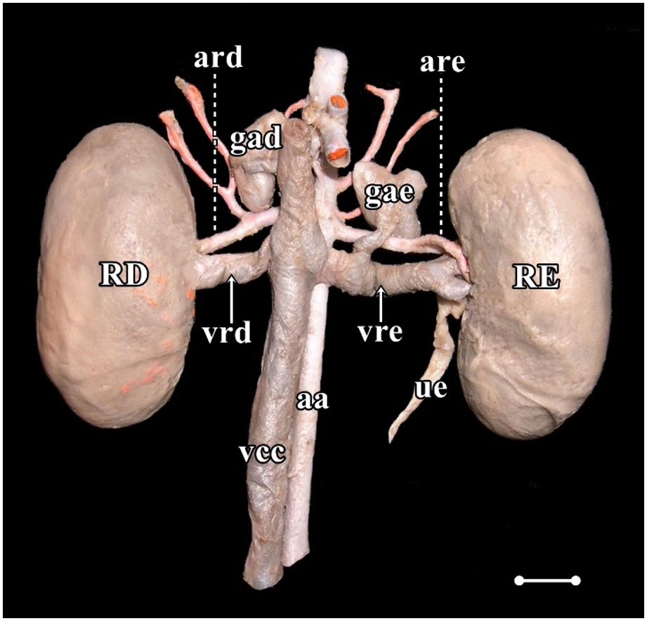
Nasua nasua is a carnivore belonging to the family Procyonidae and is widely distributed throughout South America. The details of its anatomy are fundamental to the application of antomy and understanding of its natural history. This study aimed to measure the average length, width, thickness, and volume of the kidneys; the average length of the renal artery and vein, and to describe the renal and vessel skeletopy in Nasua nasua. For this purpose, 32 kidneys and renal vessels of 16 cadaveric specimens (eight male and eight female) were dissected and measured using a digital caliper. Pearson's correlation coefficients were calculated between the rostrum-sacral length and the renal and vessel variables. The kidneys had a "bean-shaped" aspect with smooth surfaces and were unipapilate. On average, the N. nasua kidneys measured 30 × 16 × 13 mm, with no significant difference between the sexes. The average right renal artery (1.74 ± 0.67 cm) was longer than the left (1.26 ± 0.43 cm), and the right renal vein (1.22 ± 0.34 cm) was shorter than the left renal vein (1.82 ± 0.46 cm) (p < 0.05). One male animal (6.8%) presented with a double right renal vein as an anatomical variation. Both the right and left kidney cranial poles prevailed at the level of the L2 vertebra, assuming a practically symmetrical position. There was a positive and moderate to high correlation between rostrum-sacral length and renal dimensions and renal vessel lengths. The present data may be useful for interpreting the diagnosis of nephropathies that affect renal dimensions in this species and may contribute to the comparative anatomy of carnivorans.
Keywords: Neotropical carnivorans; Nephrology; South American coati; Vascularization; Wild carnivores
Downloads
References
Assunção MPB, Oliveira TAD, Oliveira, TS, Oliveira, LP, Silva DCO, Barros RAC, Silva Z. Comparative anatomy of abdominal aorta in coati (Nasua nasua). International Journal of Advanced Engineering Research and Science. 2019;6:259-267. Disponível em: https://doi.org/10.22161/ijaers.6.2.32
Trovati GR, Brito BA, Duarte JMB. Habitat use and home range of brown-nosed coati. (Nasua nasua) Carnivora: Procyonidae in the brazilian cerrado biome. Revista de Biologia Tropical. 2010;58:1069–1077.
Santos VA, Beisiegel BM. A dieta de Nasua nasua (Linnaeus. 1766) no Parque Ecológico do Tietê. SP. Revista Brasileira de Zoociências. 2006; 8:195-198.
Dyce KM, Sack WO, Wersing CJG. Tratado de Anatomia Veterinária, 5th edition. Rio de Janeiro: Elsevier, 2019.
Evans HE, DeLahunta A. Miller's Anatomy of the Dog. 4th edition. St Louis (MO): Saunders Elsevier, 2013.
Nickel RA, Schummer E, Seiferle. The anatomy of the domestic animals Berlin: Verlag Paul Parey, 1981.
Elkin. M. Kidney size. In: _____ (Ed.). Radiology of the urinary system. Boston: Little. Brown and Company, 1980. p.1014-1032.
Moell. H. Size of normal kidneys. Acta radiologica. 1956;46:640-5.
Sampaio FJ. Theoretical kidney volume versus real kidney volume: comparative evaluation in fetuses. Surgical and Radiologic Anatomy. 1995;17:71-75. Disponível em:: https://doi.org/10.1007/bf01629504
Souza EC, Leao Neto LF, Santos EAR, Abidu-Figueiredo M, Carvalho AD, Souza Junior P. Caracterización anatómica de los riñones del Zorro Pampeano (Lycalopex gymnocercus): morfometría y vasos renales. Revista Electrónica de Veterinaria. 2018;19:1-8.
Souza-Junior P, Souza EC, Viana-Peçanha S, Bernardes FCS, Montana MM, Abidu-Figueiredo M. Dimensions and skeletopy of kidneys in two populations of Cerdocyon thous (Linnaeus, 1766). Acta Veterinaria Brasílica. 2020;14:106-114. Disponível em: https://doi.org/10.21708/avb.2020.14.2.9126
Stocco AV, Sousa CAS, Gomes MS, Souza Junior P, Abidu-Figueiredo M. Is there a difference between the right and left kidney? A macroscopic approach in Brazilian Shorthair Cat. Arquivo Brasileiro de Medicina Veterinária e Zootecnia. 2016;68:1137-1144. Disponível em: https://doi.org/10.1590/1678-4162-8339
Evans H, An NQ. Anatomy of the ferret. In: Fox JG, Marini RP. Biology and Diseases of the Ferret. 3rd edition. Oxford (UK): Wiley Blackwell, 2014. p. 23–68.
Moorthy HK, Venugopal P. Measurement of renal dimensions in vivo: A critical appraisal. Indian Journal of Urology. 2011;27:169-175. Disponível em: https://doi.org/10.4103/0970-1591.82832
Milanelo L, Moreira MB, Fitorra LS, Petri BSS, Alves M, Santos AC. Occurrence of parasitism by Dioctophyma renale in ring-tailed coatis (Nasua nasua) of the Tiete Ecological Park, São Paulo, Brazil. Pesquisa Veterinaria Brasileira. 2009;29:959-962. Disponível em: https://doi.org/10.1590/S0100-736X2009001200001
Ferro BS, Hippólito AG, Castiglioni MCR, Junior JIDSS, Teixeira CR, Gonçalves RAB, Melchert A. Medidas biométricas, avaliação do escore corporal e índice de massa corpórea em Quatis (Nasua nasua) de vida livre da região Centro Sul do estado de São Paulo, Brasil. Acta Scentiae Veterinariae. 2019;47: 1639-45.
Ribeiro RG, Costa APA, Bragato N, Fonseca AM, Duque JCM, Prado TD, Silva ACR, Borges NC. Normal sonographic anatomy of the abdomen of coatis (Nasua nasua Linnaeus 1766). BMC Veterinary Research. 2013;9:124-134. Disponível em: https://doi.org/10.1186/1746-6148-9-124
Gompper ME, Decker DM. Nasua nasua. Mammalian Species 1998; 580: 1–9. Disponível em: https://doi.org/10.2307/3504444
Lobacz MA, Sullivan M, Mellor D, Hammond G, Labruyère J, Dennis R. Effect of breed, age, weight and gender on radiographic renal size in the dog. Veterinary Radiology and Ultrasound. 2012;53:437-441. Disponível em: https://doi.org/10.1111/j.1740-8261.2012.01937.x
Mareschal A, d'Anjou MA, Moreau M, Alexander K, Beauregard G. Ultrasonographic measurement of kidney-to-aorta ratio as a method of estimating renal size in dogs. Veterinary Radiology and Ultrasound. 2007;48:434-438. Disponível em: https://doi.org/10.1111/j.1740-8261.2007.00274.x
Sampaio KMOR, Araújo RB. Ultrassonografia de características lineares e estimativas do volume de rins de cães. Arquivo Brasileiro de Medicina Veterinária e Zootecnia. 2002;54:248-254. Disponível em: https://doi.org/10.1590/S0102-09352002000300005
Sohn J, Yun S, Lee J, Chang D, Choi M, Yoon J. Reestablishment of radiographic kidney size in Miniature Schnauzer dogs. Journal of Veterinay Medical Science. 2016;78:1805–1810. Disponível em: https://doi.org/10.1292/jvms.16-0003
Baitchman, EJ, Kollias, GV. Clinical anatomy of the North American river otter (Lontra canadensis). Journal of Zoo and Wildlife Medicine. 2000;31:473-483. Disponível em: https://doi.org/10.1638/1042-7260(2000)031[0473:caotna]2.0.co;2
Peçanha SV, Dünkel-Duarte R, Bernardes FCS, Estruc TM, do Nascimento RM, Dos Santos-Sousa CA, de Souza Junior P, Abidu-Figueiredo M. Anatomical characterization of the kidneys of Didelphis aurita (Didelphimorphia: Didelphidae). Folia Morphologica. 2020; 5:805-810. Disponível em: https://doi.org/10.5603/FM.a2020.0006
König HE, Maierl J, Liebich HG. Systema urinarium. In: König HE, Liebich HG. Veterinary Anatomy of Domestic Animals, 6th edition, New York, NY: Schattauer. 2016. p. 399-410.
Reis RH, Tepe P. Variations in the pattern of renal vessels and their relation to the type posterior vena cava in the dog (Canis familiaris). American Jounal of Anatomy. 1956; 99:1-15. Disponível em: https://doi.org/10.1002/aja.1000990102
Fagundes GM, Souza A, Borelli V, Riella ACM. Contribuição ao estudo da drenagem sanguínea do rim de cães (Canis familiaris – Linnaeus, 1758). Biotemas. 1990;3:117-127.
Campos CBA, Rocha OS, Abidu-Figueiredo M. Veia renal dupla em gatos: relato de casos. Revista Academica de Ciências Agrárias e Ambientais. 2014;12:127-131. Disponível em: https://doi.org/10.7213/academica.12.02.AO06
Stocco AV, Stocco NV, Santos-Sousa CA, Abidu-Figueiredo M. Veia renal tripla em gato: relato de casos. Revista Portuguesa de Ciências Veterinárias. 2014;109:120-122.
Stocco AV, Silva SC, Toledo KS, Sousa CAS, Carvalho RBJ, Abidu-Figueiredo M. Veia renal direita dupla em jaguatirica (Leopardus pardalis): relato de caso. Revista Portuguesa de Ciências Veterinárias. 2017;112:83-86.
Stocco AV, Silva SC, Toledo KS, Sousa CAS, Carvalho RBJ, Abidu-Figueiredo M. Duplicidade da veia renal direita em gato-do-mato- pequeno (Leopardus guttulus): relato de caso. Revista Academica de Ciência Animal. 2018;16:1-6.
Stocco A, Oliveira R, Santos-Sousa CA, Júnior P, Abidu-Figueiredo M. Duplicity of the right renal vein in Puma concolor (Carnivora: Felidae): a case report. Acta Scientiae Anatomica 2018;1:29-32.
Published
How to Cite
Issue
Section
License
Copyright (c) 2022 Brazilian Animal Science/ Ciência Animal Brasileira

This work is licensed under a Creative Commons Attribution 4.0 International License.
Authors who publish with this journal agree to the following terms:
- Authors retain copyright and grant the journal right of first publication with the work simultaneously licensed under a Creative Commons Attribution License that allows others to share the work with an acknowledgement of the work's authorship and initial publication in this journal.
- Authors are able to enter into separate, additional contractual arrangements for the non-exclusive distribution of the journal's published version of the work (e.g., post it to an institutional repository or publish it in a book), with an acknowledgement of its initial publication in this journal.
- Authors are permitted and encouraged to post their work online (e.g. in institutional repositories or on their website) prior to and during the submission process, as it can lead to productive exchanges, as well as earlier and greater citation of published work (See The Effect of Open Access).


