
Anatomy of the basal nuclei of Alouatta belzebul
DOI:
https://doi.org/10.1590/1809-6891v22e-70584Abstract
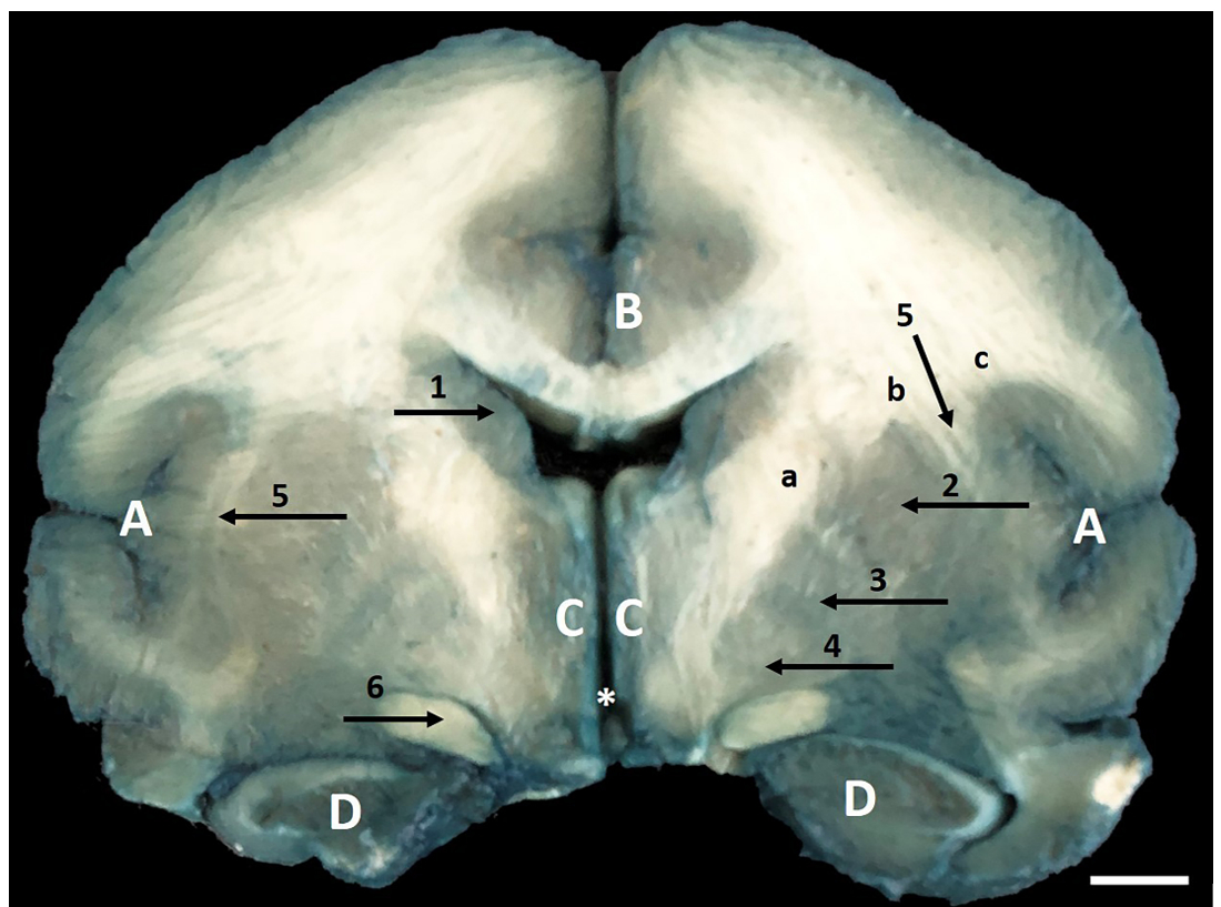
The basal nuclei are well-defined bodies of neurons with specific functions, located inside the white medullary center of the brain, directly involved with the motor system, participating greatly in the planning and control processes of movements. Studies on these nuclei in non-human primates are small and in the Alouatta belzebul species, nonexistent. The aim of the present study was to
describe the morphology of the nuclei at the base of the brain of Alouatta belzebul. Ten male and female Alouatta belzebul brains were used, where after removal and coronal cut of the brain, the Mayland technique was performed to show the basal nuclei. There was the presence of the caudate nucleus, lentiform nucleus (this formed by the putamen, medial globus pallidus and lateral globus pallidus), claustrum and substantia nigra, which, functionally, are related to motor control. The substantia nigra is part of the midbrain and is also related to learning resulting from the effects of dopamine, responsible for activating the reward and addiction system in the telbrain and is also related to the red nucleus, which is also a midbrain nucleus. In Alouatta belzebul the red nucleus is present. It was found in the literature that degeneration of substantia nigra cells can cause Parkinson's disease in Macaca fasciculares, and because Alouatta belzebul has the same anatomical structures in the basal nuclei of the base of Macaca
fasciculares, it is suggested that studies of functional evaluation of these structures should be carried out to verify whether Alouatta belzebul can be used as an experimental model for Parkinson's disease.
Keywords: Basal ganglia; Substantia nigra; Putamen; Globe pale; Howler monkey.
Downloads
References
Nascimento FF, Bonvicino CR, de Oliveira MM, Schneider MPC, Seuánez HN. Population Genetic Studies of Alouatta belzebul from the Amazonian and Atlantic Forests. American Journal of Primatology. 2008; 70: 423-431.
Valença-Montenegro, M.M., Fialho, M.S., Carvalho, A.S., Ravetta, A.L., Régis, T., de Melo, F.R., Jerusalinsky, L., Veiga, L.M., Mittermeier, R.A., Cortes-Ortíz, L. & Talebi, M. 2021. Alouatta belzebul (amended version of 2019 assessment). The IUCN Red List of Threatened Species 2021: e.T39957A190412426. https://dx.doi.org/10.2305/IUCN.UK.2021-1.RLTS.T39957A190412426.en. Downloaded on 26 September 2021.
ICMBio (Instituto Chico Mendes de Conservação da Biodiversidade). Livro Vermelho da Fauna Brasileira Ameaçada de Extinção: Volume II–Mamíferos. [http://icmbio.gov.br/portal/images/stories/comunicacao/publicacoes/publicacoes-diversas/livro_vermelho_2018_vol2.pdf].
Amado LTM, Sousa GC, Silva DCO, Silva Z, Júnior RB, Neto MAF, Lizardo FB, Santos LA, Barros RAC, Santos ALQ. Anatomia da fixação proximal do músculo reto femoral em humanos, Cebus apella e Alouatta guariba. Pubvet. 2011; 5: 1072-1078.
Leonel LCPC, Lima TC, Felipe RL, Silva EM, Silva GAO, Silva DCO, Carvalho-Barros RA, Silva Z. Anatomia descritiva da traqueia do macaco-prego (Sapajus apella). Biotemas. 2013; 26: 179-183.
Mayor P, Bowler M, Lopez APC. Functional morphology of the female genital organs in the peruvian red uakari monkey (Cacajao calvus ucayalii). Am. J. Primatol. 2013; 75: 545–554.
Lopes GP, Brito AB, Paim FP, Santos RR. Comparative characterization of the external genitalia and reproductive tubular organs of three species of the genus Saimiri Voigt, 1831 (Primates: Cebidae). Anatomia, Histologia Embryologia. 2016; 46: 143-161.
Silva EV, Silva SF, Aversi-Ferreira RAGMF, Abreu T. Nishijo H, Aversi-Ferreira TA. Comparative anatomy of the pelvic nerves in bearded capuchins (Sapajus sp). Braz. J. Vet. Res. Anim. Sci. 2016; 53: 1-17.
Teixeira DG, Hamlett WC, Guimarães MABV, Morini AC, Araújo KPC, Cury FS, Souza AF, Vidane AS, Ambrósio CE, Miglino MA. Morphological Tools for Describing the Male External Genitalia of Sapajus apella. Zoolog. Sci. 2015; 32: 97-104.
Lima AR, Guimarães SB, Branco E, Giese, EG, Muniz JAPC, Ricci REG, Miglino MA. Anatomy and histology of the urinary tract in the capuchin monkey (Sapajus apela). Pesq. Vet. Bras. 2016; 36: 221-226.
Souza-Terra DR, Sabec-Pereira DK, Lima FC, Melo FCSA, Melo FR, Pereira KF: Anatomy of the spinal cord of Alouatta belzebul. Acta Veterinaria Brasilica. 2018; 12: 55-61.
Sabec-Pereira DK, Lima FC, Melo FR, Melo FCSA, Pereira KF, Vulcani VAS: Vascularization of the Alouatta belzebul brain base. Pesq. Vet. Bras. 2020; 40: 315-323.
Segantine ACL, Melo FCSA, Melo FR, Schell RK, Zarpelon-Schutz AC, Sabec-Pereira DK, Pereira KF. Morfologia do tubo digestório de Alouatta belzebul. Research, Society and Development, 2020; 9: e5229108930.
Sabec-Pereira DK, Melo FR, Melo FCSA, Pereira KF, Vulcani VAS: Anatomy of the dura mater venous sinus of Alouatta belzebul. Anat Histol Embryol, 2020; 50: 58-64.
Pereira ER, Pires VCMC, Fernandes RJ, Sabec-Pereira DK, Melo FR, Schell RKW, Zarpelon-Schutz AC, Pereira KF. Anatomia do sistema reprodutor feminino de Alouatta belzebul (Linnaeus, 1766). Arq. Bras. Med. Vet. Zootec. 2020; 72: 2101-2110.
Prada I. Neuroanatomia funcional em medicina veterinária com correlações clínicas. Jaboticabal: Terra Molhada; 2014.
Machado ABM, Haertel LM. Neuroanatomia Funcional. São Paulo: Atheneu; 2014.
Meneses MS. Neuroanatomia aplicada. São Paulo: Grupo Gen-Guanabara Koogan; 2016.
Noureldine MHA. Fundamentos da neuroanatomia: um guia clínico. Rio de Janeiro: Elsevier; 2019.
Mainland D. Uber makroskopiche faerburg von Gehirnprraeparaten mit Berlinblau. Anat Anz. 1926; 65: 85-88.
Merini TT, Krum LK, Colman J. et al. Comparative analysis of human brain with two diferent staining techniques. UEPG Ci. Biol. Saúde. 2014; 20: 99-104.
Word Association of Veterinary Anatomists:. Nomina Anatomica Veterinaria. Ithaca: International Comittee On Veterinary Gross Anatomical Nomenclature. 2017; 35-160.
Sociedade Brasileira de Anatomia. International Anatomical Terminology. Anatomical CdT. São Paulo: Editora Manole; 2001.
Watanabe L, Madeira MC. The anatomy of the brain of the tufted capuchin (Cebus apella LINNAEUS, 1758). Rev. Odont. UNESP. 1982; 11: 5–12.
Santos JML. Estruturação de uma plataforma de ensino referente à constituição anatômica do Sistema Nervoso Central do Macaco-prego (Cebus apella). São Paulo: USP. 2019; 25-85.
Geist FD. The brain of the Rhesus monkey. Journal of Comparative Neurology, 1930; 50: 333-375.
Machado ABM. Neuroanatomia Funcional. São Paulo: Atheneu, 1993, 140-224.
Reser DH, Richardson KE, Montibeller MO, Zao S, Chan JMH, Soares JGM, Chaplin TA, Gattass R, Rosal MGP. Claustrum projections to prefrontal cortex in the capuchin monkey (Cebus apella). Frontiers in systems neuroscience, 2014; 8: 1–10.
Tilney F. The brain stem of tarsius. A critical comparison with other primates. The Journal of Comparative Neurology, 1927; 43: 371-432.
Kikuchi T, Morizane A, Doi D, Magotani H, Onoe H, Hayashi T, Mizuma H, Takara S, Takahashi R, Inoue H, Morita S, Yamamoto M, Okita K, Nakagawa M, Parmar M, Takahashi J. Human iPS cell-derived dopaminergic neurons function in a primate Parkinson’s disease model. Nature. 201; 548: 592–596.
Published
How to Cite
Issue
Section
License
Copyright (c) 2022 CIÊNCIA ANIMAL BRASILEIRA (Brazilian Animal Science)

This work is licensed under a Creative Commons Attribution 4.0 International License.
Authors who publish with this journal agree to the following terms:
- Authors retain copyright and grant the journal right of first publication with the work simultaneously licensed under a Creative Commons Attribution License that allows others to share the work with an acknowledgement of the work's authorship and initial publication in this journal.
- Authors are able to enter into separate, additional contractual arrangements for the non-exclusive distribution of the journal's published version of the work (e.g., post it to an institutional repository or publish it in a book), with an acknowledgement of its initial publication in this journal.
- Authors are permitted and encouraged to post their work online (e.g. in institutional repositories or on their website) prior to and during the submission process, as it can lead to productive exchanges, as well as earlier and greater citation of published work (See The Effect of Open Access).


