Oftalmopatias em 574 cães da raça Shih tzu atendidos em um Hospital Veterinário: estudo retrospectivo
DOI:
https://doi.org/10.1590/1809-6891v25e-79326EResumo
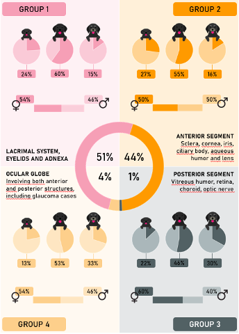
Os cães das raças braquicefálicas incluindo os Shih tzu, são predispostos ao desenvolvimento de afecções oftálmicas em razão da sua conformação facial. O objetivo principal do presente trabalho foi investigar as principais oftalmopatias em cães da raça Shih tzu atendidos no Hospital Veterinário “Governador Laudo Natel” da Universidade Estadual Paulista Júlio de Mesquita Filho, Jaboticabal- São Paulo, Brasil, entre os anos de 2014 e 2022. Foram selecionadas 574 fichas totalizando 1724 diagnósticos. Em relação sexo 50% (287/574) eram fêmeas e 50% (287/574) eram machos. Em relação à classificação anatômica os diagnósticos do grupo 1 (sistema lacrimal, pálpebras e anexos) foram os mais expressivos com 55% (945/1724), seguido do grupo 2 (segmento anterior) com 40% (683/1724), grupo 4 (bulbo ocular) com 4% (73/1724) e grupo 3 (segmento posterior) com 1% (23/1724). A idade média do diagnóstico foi de 5,2 anos, sendo que os cães adultos foram os mais representativos com 55% (317/574), seguido dos jovens 25% (145/574) e dos idosos com 20% (112/574). Os cães idosos obtiveram mais diagnósticos de glaucoma e de catarata quando comparados aos animais jovens e adultos. Em cães jovens as afecções mais diagnosticadas foram as ceratites ulcerativas e distiquíase, enquanto nos adultos e nos idosos foram ceratoconjuntivite seca e ceratite ulcerativa.
Downloads
Referências
Ekenstedt KJ, Crosse KR, Risselada M. Canine brachycephaly: anatomy, pathology, genetics and welfare. Journal of Comparative Pathology. 2020;176:109-115. Available at: https://doi.org/10.1016/j.jcpa.2020.02.008.
Packer RM, Hendricks A, Burn CC. Impact of facial conformation on canine health: corneal ulceration. PLoS One. 2015;10(5):e0123827. Available at: https://doi.org/10.1371/journal.pone.0123827.
Nutbrown-Hughes D. Brachycephalic ocular syndrome in dogs. Animal Companion. 2021;26(5):1-9. Available at: https://doi.org/10.12968/coan.2020.0056.
Castro SML, Bezerra RCF. Brazilian Cinophilia Confederation. In: Official Standard of the Shih Tzu breed. 2017.p.2-7. Portuguese. Available at: https://cbkc.org/application/views/docs/padroes/padrao-raca_200.pdf.
O'Neill DG, Lee MM, Brodbelt DC, Church DB, Sanchez RF. Corneal ulcerative disease in dogs under primary veterinary care in England: epidemiology and clinical management. Canine Genetics and Epidemiology. 2017;4:1-12. Available at: https://doi.org/10.1186/s40575-017-0045-5.
O'Neill DG, Brodbelt DC, Keddy A, Church DB, Sanchez R. Keratoconjunctivitis sicca in dogs under primary veterinary care in the UK: an epidemiological study. Journal of Small Animal Practice. 2021;62(8):636-645. Available at: https://doi.org/10.1111/jsap.13382.
Papaioannou NG, Dubielzig RR. Histopathological and immunohistochemical characteristics of vitreoretinopathy in Shih Tzu dogs. Journal of Comparative Pathology. 2013;148(2-3):230-235. Available in: https://doi.org/10.1016/j.jcpa.2012.05.014.
Kobashigawa KK, Lima TB, Padua IRM, Barros Sobrinho AAFD, Marinho FDA, Ortêncio KP, Laus JLO. Ophthalmic parameters in adult Shih Tzu dogs. Rural Science. 2015;45:1280-1285. Available in: https://doi.org/10.1590/0103-8478cr20141214.
Appelboam HP. Pug appeal: brachycephalic ocular health. Animal Companion. 2016;21(1):29-36. Available in: https://doi.org/10.12968/coan.2016.21.1.29.
Palmer SV, Gomes FE, Mcart JA. Ophthalmic disorders in a referral population of seven breeds of brachycephalic dogs: 970 cases (2008–2017). Journal of the American Veterinary Medical Association. 2021;259(11):1318-1324. Available in: https://doi.org/10.2460/javma.20.07.0388.
Sebbag L, Silva APS, Santos ÁP, Raposo ACS, Oriá AP. An eye on the Shih Tzu dog: ophthalmic examination findings and ocular surface diagnoses. Veterinary Ophthalmology. 2023;26:59-71. Available at: https://doi.org/10.1111/vop.13022.
Iwashita H, Wakaiki S, Kazama Y, Saito A. Breed prevalence of canine ulcerative keratitis according to depth of corneal involvement. Veterinary Ophthalmology. 2020;23(5):849-855. Available at: https://doi.org/10.1111/vop.12808.
Costa J, Steinmetz A, Delgado E. Clinical signs of brachycephalic ocular syndrome in 93 dogs. Irish Veterinary Journal. 2021;74(1):1-8. Available at: https://doi.org/10.1186/s13620-021-00183-5.
James-Jenks EM, Pinard CL, Charlebois PR, Monteith G. Evaluation of corneal ulcer type, skull conformation, and other risk factors in dogs: A retrospective study of 347 cases. The Canadian Veterinary Journal. 2023;64(3):225-234. Available at: https://europepmc.org/backend/ptpmcrender.fcgi?accid=PMC9979749&blobtype=pdf.
Gelatt KN, Ben-Shlomo G, Gilger BC, Hendrix DV, Kern TJ, Plummer CE. Veterinary Ophthalmology. 6th edition. Florida: John Wiley & Sons; 2021. 1082-1173p. English.
Rajaei SM, Faghihi H, Zahirinia F. The Shih Tzu eye: Ophthalmic findings of 1000 eyes. Veterinary Ophthalmology. 2024. Available at: https://doi.org/10.1111/vop.13182.
Sanchez RF, Innocent G, Mold J, Billson FM. Canine keratoconjunctivitis sicca: disease trends in a review of 229 cases. Journal of Small Animal Practice. 2007;48(4):211-217. Available at: https://doi.org/10.1111/j.1748-5827.2006.00185.x.
Deepika A, Nagaraj P, Kumar VA, Rani MU. Clinico-ophthalmic findings of corneal ulcers in dogs. International Journal of Veterinary Sciences and Animal Husbandry. 2023;8(5):220-224. Available at: https://www.veterinarypaper.com/pdf/2023/vol8issue5/PartD/8-5-25-645.pdf.
Bolzanni H, Oriá AP, Raposo ACS, Sebbag L. Aqueous tear assessment in dogs: impact of cephalic conformation, inter-test correlations, and test-retest repeatability. Veterinary Ophthalmology. 2020;23(3):534-543. Available at: https://doi.org/10.1111/vop.12751.
Gupta A, Heigle T, Pflugfelder SC. Nasolacrimal stimulation of aqueous tear production. Cornea. 1997;16(6):645-648. Available at: https://journals.lww.com/corneajrnl/citation/1997/11000/Nasolacrimal_Stimulation_of_Aqueous_Tear.8.aspx.
Gipson IK. Age-related changes and diseases of the ocular surface and cornea. Investigative Ophthalmology & Visual Science. 2013;54(14):ORSF48-ORSF53. Available at: https://doi.org/10.1167/iovs.13-12840.
Kitamura Y, Maehara S, Nakade T, Miwa Y, Arita R, Iwashita H, Saito A. Assessment of meibomian gland morphology by noncontact infrared meibography in Shih Tzu dogs with or without keratoconjunctivitis sicca. Veterinary Ophthalmology. 2019;22(6):744-750. Available at: https://doi.org/10.1111/vop.12645.
Sebbag L, Sanchez RF. The pandemic of ocular surface disease in brachycephalic dogs: The brachycephalic ocular syndrome. Veterinary Ophthalmology. 2023;26:31-46. Available at: https://doi.org/10.1111/vop.13054.
Jondeau C, Gounon M, Bourguet A, Chahory S. Epidemiology and clinical significance of canine distichiasis: A retrospective study of 291 cases. Veterinary Ophthalmology. 2023; 26(4): 339-346. Available at: https://doi.org/10.1111/vop.13091.
Yi NY, Park SA, Jeong MB, Kim MS, Lim JH, Nam TC, Seo K. Medial canthoplasty for epiphora in dogs: a retrospective study of 23 cases. Journal of the American Animal Hospital Association. 2006;42(6):435-439. Available at: https://doi.org/10.5326/0420435.
Park SA, Yi NY, Jeong MB, Kim WT, Kim SE, Chae JM, Seo KM. Clinical manifestations of cataracts in small breed dogs. Veterinary Ophthalmology. 2009;12(4):205-210. Available at: https://doi.org/10.1111/j.1463-5224.2009.00697.x.
Adkins EA, Hendrix DV. Outcomes of dogs presented for cataract evaluation: a retrospective study. Journal of the American Animal Hospital Association. 2005;41(4):235-240. Available at: https://doi.org/10.5326/0410235.
Gelatt KN, MacKay EO. Prevalence of breed-related glaucomas in pure-bred dogs in North America. Veterinary Ophthalmology. 2004;7(2):97-111. Available at: https://doi.org/10.1111/j.1463-5224.2004.04006.x.
Dubielzig RR, Ketring KL, McLellan GJ, Albert DM. Abnormalities associated with specific animal breeds. In: Veterinary Ocular Pathology: A Comparative Review. 1st ed. St. Louis: Saunders Elsevier. p.34-49. English. Available at: https://www.sciencedirect.com/book/9780702027970/veterinary-ocular-pathology.
Kanemaki N, Tchedre KT, Imayasu M, Kawarai S, Sakaguchi M, Yoshino A, Mizuki N. Dogs and humans share a common susceptibility gene SRBD1 for glaucoma risk. PLoS One. 2013;8(9):e74372. Available at: https://doi.org/10.1371/journal.pone.0074372.
Krohne SG. Use of the KOWA FC-1000 to Measure Aqueous Humor Protein and Cells in the Dog. Optica Publishing Group. 1991; MD3. Available: https://doi.org/10.1364/NAVS.1991.MD3.
Oshika T, Kato S, Hayashi K, Sawa M. Increasing of aqueous flare intensity with aging in normal human eyes. Nippon Ganka Gakkai Zasshi. 1989;93(3):358-362 . Available at: https://europepmc.org/article/med/2773720
El-Harazi SM, Ruiz RS, Feldman RM, Chuang AZ, Villanueva G. Quantitative assessment of aqueous flare: the effect of age and pupillary dilation. Ophthalmic Surgery, Lasers and Imaging Retina. 2002;33(5):379-382. Available at: https://doi.org/10.3928/1542-8877-20020901-07.
Asif SK, Kubai M, Mowat F, Dietrich U, Fentiman K, Iwabe S, Pederson S, Shap P. The Blue Book: Ocular disorders presumed to be inherited in purebred dogs. 14th edition. Idaho: American College of Veterinary Ophthalmologists, 2022; 914. Available at: https://ofa.org/wp-content/uploads/2023/06/ACVO-Blue-Book-2022.pdf.

Downloads
Publicado
Como Citar
Edição
Seção
Licença
Copyright (c) 2024 Ciência Animal Brasileira / Brazilian Animal Science

Este trabalho está licenciado sob uma licença Creative Commons Attribution 4.0 International License.
Autores que publicam nesta revista concordam com os seguintes termos:
- Autores mantém os direitos autorais e concedem à revista o direito de primeira publicação, com o trabalho simultaneamente licenciado sob a Licença Creative Commons Attribution que permite o compartilhamento do trabalho com reconhecimento da autoria e publicação inicial nesta revista.
- Autores têm autorização para assumir contratos adicionais separadamente, para distribuição não-exclusiva da versão do trabalho publicada nesta revista (ex.: publicar em repositório institucional ou como capítulo de livro), com reconhecimento de autoria e publicação inicial nesta revista.
- Autores têm permissão e são estimulados a publicar e distribuir seu trabalho online (ex.: em repositórios institucionais ou na sua página pessoal) a qualquer ponto antes ou durante o processo editorial, já que isso pode gerar alterações produtivas, bem como aumentar o impacto e a citação do trabalho publicado (Veja O Efeito do Acesso Livre).


