Abstract
Anatomical descriptions of partridges’ air sacs of the species Rhynchotus rufescens are scarce. This study aimed to evaluate the air sacs of this species. Ten cadaveric specimens of adult partridges, approximately 1 year old, were collected, and latex perfusion was used to solidify the material. The cervical air sac of a red-winged tinamou is smaller and has a more irregular conformation than other air sacs. The thoracic air sacs are symmetrical, and the cranial thoracic air sacs are smaller than the caudal ones. The abdominal air sacs are asymmetrical, and the largest ones extend themselves to the cloaca. Only one clavicular air sac was found, with three subdivisions: right, left, and medial. Additionally, right and left extrathoracic portions were found, passing under the clavicle. In one of the animals, the latexfilled humeri were found, and in three other ribs, vertebral diverticula were present. There is no clear relationship between taxonomy and biology versus the quantity and conformation of air sacs, because different animals with taxonomic proximity present differences. This study enhances species-specific anatomical knowledge of this species of partridge.
Keywords:
animal anatomy; respiratory system; wild animals; birds; partridge
Resumo
Descrições anatômicas dos sacos aéreos de perdizes da espécie Rhynchotus rufescens são escassos. Este estudo teve como objetivo avaliar os sacos aéreos desta espécie. Foram coletados dez espécimes cadavéricos de perdizes adultas, com aproximadamente 1 ano de idade, e utilizada perfusão de látex para solidificação do material. O saco aéreo cervical na perdiz de asa vermelha é menor e tem uma conformação mais irregular do que outros sacos aéreos. Os sacos aéreos torácicos são simétricos e os sacos aéreos torácicos craniais são menores que os caudais. Os sacos aéreos abdominais são assimétricos e os maiores estendem-se até a cloaca. Foi encontrado apenas um saco aéreo clavicular, com três subdivisões: direito, esquerdo e medial. Além disso, foram encontradas porções extratorácicas direita e esquerda, passando sob a clavícula. Em um dos animais foram encontrados úmeros preenchidos com látex e em outras três costelas estavam presentes divertículos vertebrais. Não há uma relação clara entre taxonomia e biologia versus quantidade e conformação dos sacos aéreos, pois diferentes animais com proximidade taxonômica apresentam diferenças. Este estudo aumenta o conhecimento anatômico específico desta espécie de perdiz.
1. Introduction
The red-winged tinamou (Rhynchotus rusfencens Temminck, 1815), also known as partridge, is a native bird of the Brazilian fauna, belonging to the order Tinaniforme, family Tinamidae(1). It is present in South America, and despite its declining population(1,2), the redwinged tinamou is classified as LC (Least concern) on the BirdLife International red list(2). Air sacs are thin-walled structures that are found throughout a bird’s body and play a crucial role in the respiratory process. They maintain a unidirectional flow of air through the lungs, which allows efficient gas exchange. Air sacs are also involved in thermoregulation, sound production, and buoyancy control in some aquatic species of birds(3,4).
The anatomical structure of air sacs in birds is unique and has evolved to meet flight demands. Birds have a total of nine air sacs, which are connected to their lungs and trachea. The air sacs are divided into four groups: cervical, cranial thoracic, caudal thoracic, and abdominal. Each group of air sacs has a specific function in the respiratory process, and they work together to allow birds to efficiently extract oxygen from the air(4). Birds do not possess a diaphragm, so they do not present any differentiation between thoracic and abdominal cavities. The respiratory system of birds is divided into lower and upper respiratory tract. The upper respiratory system is formed by the nasal cavity, larynx and trachea, whereas the lower respiratory tract is formed by the lungs and air sacs (8 or 9 depending on the species)(4).
Air sacs have already been described in the following birds: the domestic fowl (Gallus gallus)(5,6), domestic goose (Anser anser domesticus)(7), duck (Anas spp.)(8,9,10), turkey (Meleagris sp.) (11,12,13), ostrich (Struthio camelus)(14), quail (Coturnix coturnix)(15), Japanese quail (Coturnix coturnix japonica)(9), European red-winged tinamou, order Galliformes, (Alectoris graeca)(16), long-legged buzzard (Buteo rufinus)(17), European pidgeon (Columbia livia)(5,18,19), loon (Gavia immer)(20), White Cheeked Bulbul (Pycnonotus leucogenys)(21) and hooded crow (Corvus cornix)(22).
The clinical care of birds in wild animal medicine is considered common(23,24), but it must be remembered that such birds are an extremely diversified class, comprising 32 orders and more than 10,000 distinct species(1,25). Considering the scarcity of anatomical studies in partridges, the present study aimed to evaluate the anatomy of the air sacs in R. rufescens.
2. Materials and methods
Ten adults partridges (Rhynchotus rusfencens), approximately 1 year old, were used in this study and were obtained from the Department of Animal Improvement and Nutrition in the Animal Science course at UNESP – Botucatu Campus. The cadaveric material was collected after the slaughter of animals from projects in the production area, then injected with prevulcanized latex stained with blue dye (Suvinil®) and dissected for macroscopic evaluation. The process involved opening the skin in the cervical region, exposing the trachea, and making a vertical cut of approximately 1 cm to introduce a cannula for latex perfusion. The pectoral muscles and the sternum were removed by separating the costochondral joints and the shoulder girdle. The viscera were removed, and the latex mold was removed from the carcass.
The methodology adopted in this study was approved by the Committee on Ethics in the Use of Animals – CEUA of the School of Veterinary Medicine and Animal Science at the São Paulo State University (UNESP), filed under number 0030/2021. The project also obtained authorization from the Biodiversity Authorization and Information System– SISBIO, a division of the Brazilian Institute for the Environment and Renewable Natural Resources – IBAMA, for the temporary maintenance of wild vertebrates in captivity, under number 77786-1.
3. Results
R. rufescens air sacs are found very close to the viscera - liver, ventriculus and intestine - in the coelomic cavity. After removing the viscera, it is possible to visualize an anatomically organized set of abdominal, clavicular, cranial thoracic and caudal thoracic air sacs (Figure 1).
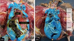
(a) Relationship of air sacs and viscera in a partridge’s coelomic cavity where the Liver, Ventriculus and Gut can be visualized (marked off by yellow lines). (b) Visualization of air sacs in a partridge’s coelomic cavity after visceral removal. Ab.: Abdominal air sac; Clav.: Clavicular air sac; Cr.T: Cranial thoracic air sac; Ca.T: Caudal thoracic air sac; Cerv.: Cervical air sac.
Partridges, commonly, do not present cervical air sacs. However, in the red-winged tinamou, we identified a total of seven air sacs, including one cervical air sac which is a smaller and more irregularly shaped cervical air sac compared to other birds. Furthermore, we observed one pair of cranial thoracic air sacs, one pair of caudal thoracic air sacs, and one pair of abdominal air sacs (Figure 2). Additionally, a clavicular air sac with three subdivisions was observed: right, left and median; there are right and left extrathoracic portions found under the clavicle (Figure 2).

(a) Visualization of a partridge’s air sacs after skin removal and ventral musculature. (b) Note the extrathoracic portion of the clavicular air sac (arrows). Ab.: Abdominal air sac; Clav.: Clavicular air sac; Cr.T: Cranial thoracic air sac; Ca.T: Caudal thoracic air sac; Cerv.: Cervical air sac.
The thoracic air sacs are symmetrical, and the cranial thoracic air sacs are smaller (approximately 2.6 cm) than the caudal ones (between 4.5 to 4.9 cm). The cranial thoracic air sacs are in close anatomical relationship with the lungs. The abdominal air sacs are asymmetrical and larger, extending to the cloaca region (Figures 3 and 4).

Latex cast of a partridge’s lower respiratory system, dorsal (a) and ventral (b) view. Ab.: Abdominal air sac; Clav.: Clavicular air sac; Cr.T: Cranial thoracic air sac; Ca.T: Caudal thoracic air sac; Cerv.: Cervical air sac; Pulm.: Lung. R=right; L=Left.

Latex cast of a partridge’s lower respiratory system, right side (above) and left side (below) view. Ab.: Abdominal air sac; Clav.: Clavicular air sac; Cr.T: Cranial thoracic air sac; Ca.T: Caudal thoracic air sac; Cerv.: Cervical air sac; Pulm.: Lung.; R=right; L=Left.
Figures 3 and 4 show latex casts of the lower respiratory system of a partridge in dorsal and ventral views, identifying the lungs and clavicular, abdominal, cranial thoracic and caudal thoracic air sacs. This view allows the understanding of the three-dimensional arrangement of these respiratory structures, providing insights into the distribution of the air sacs in relation to the vertebral column and ribs. Figure 4 shows the positioning and continuity of airways along the lateral aspect of the bird’s body. These images provide detailed representations of the avian respiratory anatomy, offering valuable insights into the study of the respiratory system adapted for flight.
Moreover, in one of the animals, both humeri and vertebral diverticula were filled with latex (Figures 5 and 6). The filling of the pneumatic cavities of a partridge’s humerus highlights the pneumatization of this bone.

A partridge’s humerus filled with latex in the pneumatic cavities, showing pneumatization of this bone.

Lateral view of a partridge’s air sacs, with visualization of latex insertion into vertebral diverticula.
The ontogeny of the birds’ pneumatic bones determines the age the bones begin to pneumatize, and the points they begin. The humerus and cervical vertebrae begin at 35 days after hatching, while the sternum only begins at 126 days after hatching.
The intervertebral diverticulum in birds is an anatomical structure located between the spinal column vertebrae, especially in the thoracic and lumbar regions. It is an extension of the air sacs, which are part of the respiratory system of birds. These projections allow air to circulate not only in the lungs, but also in some areas of the bones, such as the vertebrae, helping reduce birds’ body weight, a crucial adaptation for flight.
4. Discussion
Studies about the respiratory system of birds have been done for many years, with several manuscripts published on the topic. One study by Maina(26,27) examined the anatomy of the avian respiratory system and found that air sacs play a vital role in the respiratory process. The researchers concluded that the unique respiratory system in birds allows a more efficient extraction of oxygen from the air. Those authors obtained computational modeling to simulate the air flow in bird lungs and found that air sacs played a crucial role in the flight process(28), showing that the functional efficiency of the avian respiratory system is correlated to its structural complexity.
Birds’ ratio size of air sacs follows what has already been described in the literature for other species: the cervical air sac has a more irregular conformation and a smaller size(5,9,10,29). But, in Rhynchotus rufescens, we observed that the thoracic air sacs are symmetrical and the cranial thoracic air sacs are smaller than the caudal ones. The abdominal air sacs are the largest and, despite being asymmetrical, both antimeres extend to the cloaca. A single individual presented the right antimere smaller than the left. The left abdominal air sac was described as smaller than the right one in ducks - Anas spp(8,9,16). But this species is not phylogenetically related to R. rufescens. However, similarity between the morphological characteristics of the abdominal air sacs was observed in quails(15,29,30).
The extrathoracic portion of the clavicular air sac pneumatizes the humerus and sternum and extends to the syrinx where it plays an essential role in vocalization(3). In one of the dissected animals, the latex filling evidenced the humerus pneumatization, and in three other ribs, it revealed the presence of vertebral diverticula. Paradoxically, a study with the same technique performed on Japanese quails, which are phylogenetically close to partridges, showed that the humerus was a non-aerated bone(29).
This connection between air sacs and bone pneumatization, such as that of the humerus, one of the main bones of the wings, is an example of the functional coadaptation for flight in birds because it makes the bones lighter, reducing the total weight without compromising structural resistance. However, partridges are terrestrial birds that can fly very short distances and at low altitudes, so the permanence of this pneumatization needs to be further investigated in R. rufescens.
Latex viscosity means that pneumatized bones are not necessarily filled, and bone pneumatization is studied via a thorough osteological examination to locate diverticula and visualization of trabeculae through sectioning of the bones. Due to the fact that the present study did not focus on the description of pneumatic bones, it was limited to reporting describable bones by latex filling; therefore, it is not possible to affirm whether other bones are pneumatized in the species and whether there is an intraspecific variation.
There are no studies that corroborate or refute the correlation between the number of air sacs and phylogeny in birds, since species with taxonomic proximity present differences, as exemplified by quails, which do not have a cervical sac and have a single clavicular sac, totaling seven air sacs(30). Other species are phylogenetically distant from these and have two cervical sacs and one clavicular sac(31) ,such as ostriches (Struthio camelus), or two pairs of cervical sacs(32) such as hooded crows (Corvus cornix), totaling nine in both species when added to the other air sacs.
The morphology of the respiratory system found in the red-winged tinamou (R. rufescens) is scarcely described in the literature and is only similar to the morphology reported in loons (Gavia immer), a water bird belonging to the Gaviiformes order(20,33). The other birds with seven air sacs are geese (Anser anser domesticus)(1,7) and turkeys (genus Meleagris) with a single cervicoclavicular air sac and the other paired ones(11,12).
5. Conclusion
Red-winged tinamou (Rhynchotus rusfencens Temminck, 1815) presents a total of seven air sacs, differentiated into a single clavicular air sac, a pair of cranial thoracic air sacs, a pair of caudal air sacs, and a pair of abdominal air sacs. The ongoing research in this area is helping increase our understanding about the respiratory system of birds and its unique adaptations. These results will be important as a basis for future phylogenetic studies and for comparisons of this species with other birds.
Acknowledgments
The authors would like to thank the School of Veterinary Medicine and Animal Science at the São Paulo State University (UNESP), for all the support in this study.
References
-
1 Cubas ZS, Silva JCR, Catão-Dias JL. Tratado de Animais Selvagens. 2. ed. São Paulo: Roca; 2014. 272–353 p. Available from: https://app.minhabiblioteca.com.br/#/books/978-85-277-2649-8/
» https://app.minhabiblioteca.com.br/#/books/978-85-277-2649-8/ -
2 BirdLife International. BirdLife International. 2022 [cited 2023 Mar 20]. Red-winged Tinamou (Rhynchotus rufescens) - BirdLife species factsheet. Available from: http://datazone.birdlife.org/species/factsheet/red-wingedtinamou-rhynchotus-rufescens
» http://datazone.birdlife.org/species/factsheet/red-wingedtinamou-rhynchotus-rufescens -
3 O’Malley B. Clinical Anatomy and Physiology of Exotic Species. Philadelphia, PA: Elsevier Saunders; 2005. 113–116 p. Available from: https://doi.org/10.1016/B978-0-7020-2782-6.X5001-7
» https://doi.org/10.1016/B978-0-7020-2782-6.X5001-7 -
4 Casteleyn C, Cornillie P, Van Cruchten S, Van den Broeck W, Van Ginneken C, Simoens P. Anatomy of the lower respiratory tract in domestic birds, with emphasis on respiration. Anat Histol Embryol. 2018;47(2):89–99. Available from: https://pubmed.ncbi.nlm.nih.gov/29250822/
» https://pubmed.ncbi.nlm.nih.gov/29250822/ -
5 Akester AR. The comparative anatomy of the respiratory pathways in the domestic fowl (Gallus domesticus), pigeon (Columba livia) and domestic duck (Anas platyrhyncha). J Anat. 1960 Oct;94(Pt 4):487. Available from: https://www.ncbi.nlm.nih.gov/pmc/articles/PMC1244348/
» https://www.ncbi.nlm.nih.gov/pmc/articles/PMC1244348/ -
6 Goodchild WM. Differentiation of the body cavities and air sacs of Gallus domesticus post mortem and their location in vivo. Br Poult Sci. 1970 Apr 1;11(2):209–15. Available from: https://pubmed.ncbi.nlm.nih.gov/5420476/
» https://pubmed.ncbi.nlm.nih.gov/5420476/ -
7 Onuk B, Haziroǧlu RM, Kabak M. Gross anatomy of the respiratory system in goose (Anser anser domesticus): Bronchi and sacci pneumatici. Ankara Universitesi Veteriner Fakultesi Dergisi. 2009;56(3):165–70. Available from: http://vetjournal.ankara.edu.tr/en/
» http://vetjournal.ankara.edu.tr/en/ -
8 Araújo JM de, Roberto J, Junior K, Kastein P, Da F, Bianchi C, et al. Descrição anatômica topográfica dos sacos aéreos de patos, Anas platyrhynchos (Aves: Anseriformes). Bioscience Journal. 2014 Mar 24;30(3):810–5. Available from: https://seer.ufu.br/index.php/biosciencejournal/article/view/19817
» https://seer.ufu.br/index.php/biosciencejournal/article/view/19817 -
9 Çevik-Demirkan A, Haziroğlu RM, Kürtül İ. Air sacs (Sacci pneumatici) in mallard ducks (Anas platyrhynchos). Ankara Üniv Vet Fak Derg. 2006;53:75–8. Available from: http://vetjournal.ankara.edu.tr/tr/download/article-file/776412
» http://vetjournal.ankara.edu.tr/tr/download/article-file/776412 -
10 El-Bably SH, Rezk HM, Tolba AR. Gross morphological studies on the air sacs (Sacci pneumatici) of Golden Pekin duck (Anas platyrhyncha). Haryana Veterinarian. 2014;53(1):13–7. Available from: https://www.luvas.edu.in/haryana-veterinarian/download/harvet2014/3.pdf
» https://www.luvas.edu.in/haryana-veterinarian/download/harvet2014/3.pdf -
11 King AS, Atherton JD. The identity of the air sacs of the turkey (Meleagris gallopavo). Acta Anat (Basel). 1970;77(1):78–91. Available from: https://pubmed.ncbi.nlm.nih.gov/5504208/
» https://pubmed.ncbi.nlm.nih.gov/5504208/ -
12 Ragab SA, Reem RT. Macroscopical anatomy of the air sacs of the turkey. International Journal of Advanced Research in Biological Sciences. 2016;3(8):149–59. Available from: http://s-o-i.org/1.15/ijarbs-2016-3-8-25
» http://s-o-i.org/1.15/ijarbs-2016-3-8-25 -
13 Rigdon RH, Ferguson TM, Feldman GL, Couch JR. Air Sacs in the Turkey. Poult Sci. 1958;37(1):53–60. Available from: https://doi.org/10.3382/ps.0370053
» https://doi.org/10.3382/ps.0370053 -
14 Deeming DC. The ostrich: biology, production and health. Wallingford (United Kingdom) and New York: CABI Publishing; 1999. 13–51 p. Available from: https://lccn.loc.gov/99017680
» https://lccn.loc.gov/99017680 -
15 Bianchi P, Silvestre T, Junior JRK, Poscai AN, Leandro RM, Gonçalez PO. Relações topográficas dos sacos aéreos de codornas (Coturnix coturnix). Ciência Animal Brasileira / Brazilian Animal Science. 2016;17(2):279–84. Available from: https://doi.org/10.1590/1089-6891v17i231635
» https://doi.org/10.1590/1089-6891v17i231635 -
16 Kürtül I, Aslan K, Aksoy G, Özcan S. Morphology of the air sacs (sacci pneumatici) in the rock partridge (Alectoris graeca). Vet Res Commun. 2004;28(7):553–9. Available from: https://doi.org/10.1023/B:VERC.0000042871.69978.f0
» https://doi.org/10.1023/B:VERC.0000042871.69978.f0 -
17 Orhan İÖ, Kabak M, Oto Ç, Haziroğlu RM. Air sacs (sacci pneumatici) in the long-legged buzzard (Buteo rufinus). Ankara Üniv Vet Fak Derg. 2009;56:7–11. Available from: https://doi.org/10.1501/Vetfak_0000002180
» https://doi.org/10.1501/Vetfak_0000002180 -
18 Gilbert PW. The avian lung and air-sac system. Auk. 1939;56(1):57–63. Available from: https://doi.org/10.2307/4078361
» https://doi.org/10.2307/4078361 -
19 Müller B. Air sacs of the pigeon. Smithsonian Miscellaneous Collections. 1908;50:365–414. Available from: https://repository.si.edu/bitstream/handle/10088/23319/SMC_50_Muller_1908_28_365-414.pdf
» https://repository.si.edu/bitstream/handle/10088/23319/SMC_50_Muller_1908_28_365-414.pdf -
20 Gier HT. The air sacs of the loo. Auk. 1952;69(1):40–9. Available from: https://doi.org/10.2307/4081291
» https://doi.org/10.2307/4081291 -
21 Jaifar SK, Sawad AA. Morphological study of air sacs (Sacci pneumatici) in white cheeked bulbul (Pycnonotus leucotis). Basrah Journal of Veterinary Research. 2016 Jul 25;15(4):185–93. Available from: https://www.cabidigitallibrary.org/doi/full/10.5555/20173048471
» https://www.cabidigitallibrary.org/doi/full/10.5555/20173048471 -
22 El-Sayed AK, Hassan S. Gross morphological features of the air sacs of the hooded crow (Corvus cornix). Anat Histol Embryol. 2020 Mar 1;49(2):159–66. Available from: https://doi.org/10.1111/ahe.12504
» https://doi.org/10.1111/ahe.12504 -
23 Capaverde-Jr UD, Lopes MD, Almeida NCV, Pathek DB. Wild animals collected by the Independent Company of Environmental Police Monte Roraima in urban area of Boa Vista. Brazilian Amazon Biota Amazônia. 2018;8(1):43–8. Available from: https://www.oasisbr.ibict.br/vufind/Record/UNIFAP-3_013168198218644e05ffa7f7cd167114
» https://www.oasisbr.ibict.br/vufind/Record/UNIFAP-3_013168198218644e05ffa7f7cd167114 -
24 Lima RA, Silva SM. Survey of wild fauna in rehabilitation centre of environment military police battalion of 2010, 2011 and 2013 in the municipality of Candeias Jamari-RO. Revista Eletrônica em Gestão, Educação e Tecnologia Ambiental. 2014 Apr 15;18(1):296–311. Available from: https://doi.org/10.5902/2236117012289
» https://doi.org/10.5902/2236117012289 -
25 Tully TN, Dorrestein GM, Jones AK. Clínica de Aves. 2. ed. São Paulo: Elsevier Ltd; 2010. 150 p. Available from: https://books.google.com.br/books/about/Cl%C3%ADnica_de_aves.html?id=YtCHmVISAKkC&redir_esc=y
» https://books.google.com.br/books/about/Cl%C3%ADnica_de_aves.html?id=YtCHmVISAKkC&redir_esc=y -
26 Maina JN. Development, structure, and function of a novel respiratory organ, the lung-air sac system of birds: to go where no other vertebrate has gone. Biol Rev Camb Philos Soc. 2006 Nov;81(4):545–79. Available from: https://doi.org/10.1016/j.resp.2006.05.005
» https://doi.org/10.1016/j.resp.2006.05.005 -
27 Maina JN. Spectacularly robust! Tensegrity principle explains the mechanical strength of the avian lung. Respir Physiol Neurobiol. 2007 Jan 15;155(1):1–10. Available from: https://doi.org/10.1016/j.resp.2006.05.005
» https://doi.org/10.1016/j.resp.2006.05.005 -
28 Maina JN. Perspectives on the Structure and Function of the Avian Respiratory System: Functional Efficiency Built on Structural Complexity. Frontiers in Animal Science. 2022 Apr 13;3:851574. Available from: https://doi.org/10.3389/fanim.2022.851574
» https://doi.org/10.3389/fanim.2022.851574 -
29 Çevik-Demirkan A, Kürtül I, Haziroǧlu RM. Gross morphological features of the lung and air sac in the Japanese quail. J Vet Med Sci. 2006 Sep;68(9):909–13. https://doi.org/10.1292/jvms.68.909
» https://doi.org/10.1292/jvms.68.909 -
30 Sawad AA, Udah, AD. Morphological and histopathological study of air sacs (Sacci pneumatic) in Japanese quail (Coturnix coturnix japonica). MRVSA. 2012; 1(1), 50-56, 2012. Available from: https://doi.org/10.22428/mrvsa.2307-8073.2012.00117.x
» https://doi.org/10.22428/mrvsa.2307-8073.2012.00117.x -
31 Bezuidenhout AJ, Groenewald HB, Soley JT. An anatomical study of the respiratory air sacs in ostriches. Onderstepoort J Vet Res. 1999;66(4):317–25. Available from: https://pubmed.ncbi.nlm.nih.gov/10689704/
» https://pubmed.ncbi.nlm.nih.gov/10689704/ -
32 El-Sayed AK, Hassan S. Gross morphological features of the air sacs of the hooded crow (Corvus cornix). Anat Histol Embryol. 2020 Mar 1 [cited 2023 Jul 12];49(2):159–66. Available from: https://pubmed.ncbi.nlm.nih.gov/31617250/
» https://pubmed.ncbi.nlm.nih.gov/31617250/ -
33 BirdLife International. BirdLife International. 2020. Alectoris graeca (Rock Partridge). Available from: https://www.iucnredlist.org/species/22678684/183062253
» https://www.iucnredlist.org/species/22678684/183062253


