
Abstract
Captive Psittaciformes may harbor Gram-negative bacteria in their
digestive tract, mainly due to poor hygienic conditions and confinement.
The present study was carried out with the objective of isolating and
identifying Escherichia coli in samples collected from
Psittaciformes cages in 50 commercial establishments in the metropolitan
region of Goiania, with subsequent antimicrobial susceptibility testing
and detection of virulence genes. A total of 141 samples of excreta and
swab samples from feeders and water bowls were collected, totaling 423
samples. Escherichia coli was isolated from 9.7% (41/423)
samples: 12% (17/141) in excreta, 8.5% (12/141) in feed, and 8.5% (12
/141) in waterers. To determine the susceptibility profile of E.
coli isolates, resistance to ciprofloxacin 4.9% (2/41),
gentamicin 17.0% (7/41), doxycycline 34.1% (14/41), florfenicol 34.1%
(14/41), trimethoprim 39.0% (16/41), tetracycline 41.5% (17/41),
enrofloxacin 43.9% (18/41), amoxicillin 48.8% (20/41), neomycin 61.0%
(25/41), and sulfonamide 90.2% (37/41) was determined. In 20 isolates,
resistance was determined at 4 or more antimicrobials, seven of excreta
(7/17), five of feed (5/12), and eight of waterers (8/12). One of the
isolates from the waterers showed resistance to all antimicrobials. The
iss gene was detected in three isolates, the tsh gene in three, the papC
gene in two, traT and eae genes were not detected. In this study, it can
be concluded that Psittaciformes commercialized as pet are carry E.
coli isolates resistant to most commonly used antimicrobials,
mainly sulfonamides and neomycin, besides having virulence and serum
resistance genes, which highlights the possibility of the to cause
disease in humans.
Key words: APEC, multidrug resistance, virulence genes,
wild birds, public health
Resumo
Psittaciformes em cativeiro podem abrigar bactérias Gram-negativas em
seu trato digestivo, principalmente devido a condições higiênicas
inadequadas e ao confinamento. O presente estudo teve o objetivo de
isolar e identificar Escherichia coli em amostras coletadas de
gaiolas de Psittaciformes em 50 estabelecimentos comerciais da região
metropolitana de Goiânia, com subsequentes testes de susceptibilidade
antimicrobiana e detecção de genes de virulência. Foram coletadas 141
amostras de excrementos e suabes de alimentadores e bebedouros,
totalizando 423 amostras. Escherichia coli foi isolada em 9,7%
(41/423) amostras: 12% (17/141) em excrementos, 8,5% (12/141) em ração e
8,5% (12/141) em bebedouros. Os isolados de E. coli mostraram
resistência à ciprofloxacina 4,9% (2/41), gentamicina 17,0% (7/41),
doxiciclina 34,1% (14/41), florfenicol 34,1% (14/41), trimetoprim 39,0%
(16/41), tetraciclina 41,5% (17/41), enrofloxacina 43,9% (18/41),
amoxicilina 48,8% (20/41), neomicina 61,0% (25/41) e sulfonamida 90,2%
(37 / 41) foi determinado. Multirresistência (resistência a quatro ou
mais antimicrobianos) foi encontrada em 20 amostras, sete de excrementos
(7/17), cinco de ração (5/12) e oito de bebedouros (8/12). Um dos
isolados dos bebedouros apresentou resistência a todos os
antimicrobianos. O gene iss foi detectado em três isolados, o gene tsh
em três, o gene papC em dois, os genes traT e eae não foram detectados.
Neste estudo, pode-se concluir que os Psittaciformes comercializados
como animais de estimação são portadores de isolados de E. coli
resistentes aos antimicrobianos mais utilizados, principalmente
sulfonamidas e neomicina, além de possuir genes de virulência e
resistência sérica, destacando a possibilidade de causar doenças em
humanos.
Palavras chave: APEC, amostras multirresistentes, genes
de virulência, aves silvestres, saúde pública
Section: Medicina Veterinária
Received
September 17, 2019.
Accepted
June 1, 2020.
Published
September 18, 2020.
www.revistas.ufg.br/vet
visit the website to get the how to cite in the article page.
Introduction
Psittaciformes are commercialized as pets around the world, due to their natural characteristics(1), being beautiful colors and exotic birds. In their natural habitat, the microbiota in their digestive tract is mainly composed of Gram-positive bacteria(2,3). However, when kept as pets, which spend most of their time in limited microenvironments, the risk of exposure to potentially pathogenic bacteria becomes greater. Inadequate hygiene of the environment and containers in which water and food are supplied(4) favor the growth of Gram-negative bacteria(5,6). The risk of exposure becomes even greater in commercial establishments, where there is usually overcrowding of cages with insufficient hygiene and high stress conditions(7). According to Marietto-Gonçalves et al.(8), monitoring the presence of Gram-negative bacteria in the enteric microbiota of Psittaciformes should be included in the routine breeding of these birds; since they are not part of the physiological microbiota, there are risks of dissemination of possible pathogens to humans and other animals.
Of the Gram-negative bacteria isolated in psitacídeos(6), Escherichia coli has the highest incidence. This enterobacterium may be present in nature and even colonize the gastrointestinal tract (GIT) of birds, in a non-pathogenic way, being considered as commensal and opportunistic(9). However, some strains have modified their antigenic structures and acquired genes that have made them capable of generating diseases(10,11).
E. coli is a microorganism whose wild-type phenotype has no intrinsic resistance, and all resistances are acquired. Currently, antibiograms for E. coli have become essential considering the ecological, genetic, and environmental factors and the fact that more isolates are resistant to most antibiotics(9). According to the World Health Organization(12), so countries need to unite to implement measures and monitor studies to better understand infections, concentrating their actions in precise control and diagnostic measures for the rational use of antimicrobials.
Regarding poultry and wild birds, APEC (Avian Pathogenic E. coli) is the pathotype of E. coli(13), and its strains possess a wide genetic diversity, with a wide range of virulence factors. Among the genes that classify a strain as APEC are those responsible for resistance to the bactericidal effects of serum (traT and iss), adhesion mechanisms (pap), aerobactin (iuc) and temperature-sensitive hemagglutinin (tsh)(9, 14-16).
Studies of E. coli, isolated from humans and animals, have demonstrated the presence of several genes that it has in common indicates the possibility of genetic changes between different strains when they come in contact, contributing to a greater virulence and to the development of resistant pathogenic strains(17). Moreover, subsequent studies have pointed out an exponential increase in antimicrobial resistant microorganisms(18), with Psittaciformes being the focus of some studies that have revealed their importance as hosts of bacteria resistant to antibiotics(19)
Considering the importance of the investigation of bacterial strains present in commercially-reared animals as pets, which may represent a challenge in the epidemiological control of the human-animal relationship, the present study aimed to identify the presence of E. coli in samples collected in Psittaciformes' cages, with the evaluation of the antimicrobial resistance profile more commonly employed to treat these birds therapeutically and detection of virulence genes in bacterial isolates.
Material and methods
The present study was approved by the Commission of Ethics in the Use of Animals of the Federal University of Goiás (CEUA -UFG) under the protocol nº 058/17.
After updating the official stores register, together with the governmental inspection department, establishments with the presence of birds (Psittaciformes) were identified metropolitan regions of Goiânia-Goiás-Brazil in the second half of 2017, and then 50 establishments were selected to collect samples. The selection of the number of cages for sample collection was established according to the total number of cages containing per establishment. In each selected cage, samples were collected from excreta, feeders and waterers, in a total of three samples per cage.
Approximately 1.0 g of excreta were collected at five different points from the cage trays, thus forming a pool, which constituted a sample. From the feeders, 2.5 g of feed were collected in five different points, constituting a sample. The waterers were swabbed all over the surface three times, which constituted a sample. After collection, the samples were homogenized, identified and transported in isothermal boxes containing ice to the Laboratory of Bacteriology and Molecular Diagnostics of the Department of Veterinary Medicine (EVZ-UFG) for processing. At the end of the collection in the 50 establishments, a total of 423 samples were obtained, 141 from each source (feeder, waterers and excreta).
Eight different species of Psittaciformes were identified, distributed in the 141 cages of the 50 studied commercial establishments: budgerigar (Melopsittacus undulatus) in 48.2% (68/141); cockatiels (Nymphicus hollandicus) in 33.4% (47/141); peach-faced loverbirds (Agapornis roseicollis) in 14.2% (20/141); bourke's parrots (Neopsephotus bourkii) in 1.4% (2/141); red rumped parrots (Psephotus haematonotus) in 0.7% (1/141); turquoise-fronted parrot (Neophema pulchella) in 0.7% (1/141); blue-fronted amazon parrot (Amazona aestiva) in 0.7% (1/141) and scarlet macaw (Ara macao macao) in 0.7% (1/141). It was found that most of the cages contained more than two birds (100/141).
The samples were processed according to Oliveira(20). Initially, the feed samples were weighed and, like the waterer swab samples, were processed in 1% peptone water, at 1:10, and incubated at 37 °C for 18-24 h. After, 1 mL of this solution was transferred to 9 mL of cystine selenite broth (CS) and then incubated at 37 °C for 18-24 h. The excreta samples were weighed and inoculated in brain heart infusion (BHI) and incubated at 37 °C for 18-24 h.
After incubation, CS and BHI broths aliquots were streaked with a nickel loop on MacConkey agar and incubated at 37 °C for 18-24 h. Three colony forming units (CFU) with morphological characteristics of Escherichia coli were transferred to tubes containing triple sugar iron agar (TSI) and incubated at 37 °C for 24 h.
After that period, the TSI tubes were selected according to the use of glucose, sucrose and lactose and submitted to tests of motility, indole production, urease production, H2S production, malonate, methyl red reaction, and Simmons citrate. The isolates with compatible characteristics of E. coli were spiked in BHI broth, incubated at 37 °C for 24 h and kept at -20 °C for later use.
The antimicrobial susceptibility profile of the E. coli isolates was determined by the Disk Diffusion method according to Clinical and Laboratory Standards Institute - CLSI(21). Antimicrobials tested were: amoxicillin (10 µg), gentamicin (10 µg), ciprofloxacin (5 µg), enrofloxacin (5 µg), florfenicol (30 µg), neomycin (30 µg), sulfonamide 300 µg), tetracycline (30 µg), trimethoprim (25 µg) and doxycycline (30 µg). E. coli ATCC 25922 was used as a reference isolate.
For DNA extraction, the Qiagen Plasmid mini kit and Wizard™ Genomic DNA Purification kit were used, and their protocols were followed using 3 mL of suspension of the bacterial culture in BHI broth, incubated for 24 h at 37 °C. The DNA pellets obtained on extraction were suspended in 50 μL of buffer (TE). The buffer solution was prepared as 242 g TRIS 2 M base (2-amino-2-hydroxymethyl-propane-1,3-diol), 57.1 mL glacial acetic acid, 100 mL disodium EDTA solution (Na2 EDTA) 0.5 M (pH 8.0), and distilled water to one liter (v:v), and then stored at -20 °C. For DNA amplification, the isolates were submitted to standard PCR using different primers for the detection of the following genes: tsh (5'-ACT ATT CTC TGC AGG AAG TC-3'; 5'-CTT CCG ATG TTC TGA ACG T-3'; 829 bp)(16), iss (5'-ATC ACA TAG GAT TCT GCC G-3'; 5'-CAG CGG AGT ATA GAT GCC A-3'; 309 bp)(16), traT (5'-GGT GTG GTG CGA TGA GCA CAG-3';5'-CAC GGT TCA GCC ATC CCT GAG-3'; 290 bp)(22), papC (5'-TGA TAT CAC GCA GTC AGT AGC-3'; 5'-CCG GCC ATA TTC ACA TAA-3'; 205 bp)(23), and eae (5'-AAA CAG GTG AAA CTG TTG CC-3';5'-CTC TGC AGA TTA ACC TCT GC-3'; 454 bp)(24).
For iss, traT, papC and eae gene analysis, a polymerase chain reaction was performed in a thermocycler at a temperature of 90 °C for five minutes and 30 cycles of amplification, comprised by denaturation at 94 °C for one minute, 50 °C for one minute and extension at 72 °C for two minutes. This was followed by a final step at 72 °C for seven minutes.
The amplified genes were subjected to electrophoresis on 0.8% agarose gel at 90 V for a period of 50 minutes. The agarose gel was prepared with 0.8 mg of agarose, 10 mL of TEB (0.5X), 100 mL of double distilled water (q.s.p). TEB buffer was prepared with 5.4 g of Tris base, 2.0 mL of 0.5 M EDTA (pH 8.8), 2.75 g of boric acid, and 100 mL of double distilled water (q.s.p). The gel was stained with GelRed (Uniscience™) and visualized under ultraviolet light on a transilluminator.
Results
Among the 423 samples analyzed, E. coli was isolated from 12% (17/141 isolates) from excrement, 8.5% (12/141 isolates) from feed and 8.5% (12/141 isolates) from waterers swabs.
In the determination of the susceptibility of E. coli isolates from excreta, feed and waterers, a higher resistance to the antimicrobials sulfonamide and neomycin was observed (Figure 1).
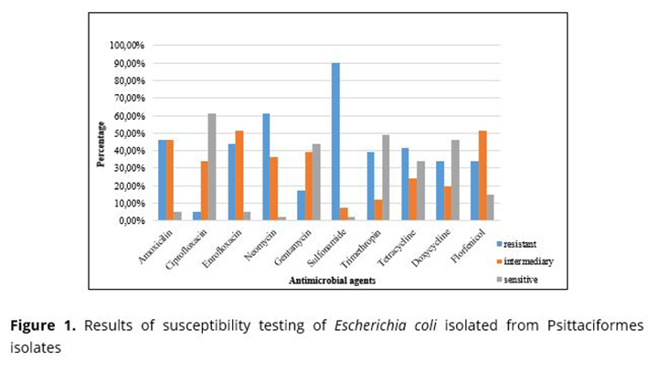
Among the isolates from waterers, all were resistant to sulfonamide (12/12). In isolates from feed and excreta, there was no resistance to ciprofloxacin.
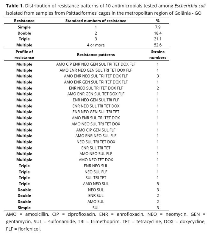
Among the 41 E. coli isolates from the samples obtained from the Psittaciformes' cages, multi-resistance (resistance to four or more antimicrobials) was found in 20 samples, seven of excreta (7/17), five of feed (5/12), and eight of waterers (8/12). One of the isolates from the waterers showed resistance to all antimicrobials (Table 1).
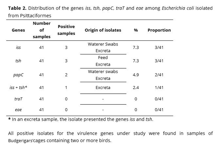
As for the virulence genes, three isolates of E. coli with the iss gene (7.3%) were detected: two from excreta and one from feed samples; three isolates with the tsh gene (7.3%) were found: two from feed and one from excreta. Among these isolates, one from excreta presented both genes (tsh and iss). The papC gene was also found in two isolates, one from a waterer sample and the other from an excreta sample. The traT and eae genes were not found in any isolate (Table 2).
All positive isolates for the virulence genes under study were found in samples of Budgerigars'cages containing two or more birds.
Discussion
The presence of E. coli, isolated from the excreta, does not characterize enteric disturbance or disease in the animal, since the birds were apparently healthy. The results obtained in the research coincide with the reports of other authors, who reported that E. coli can be detected in clinically healthy birds acting as an opportunistic bacterium that generates clinical disease only in immunosuppressed birds subjected to high stress loads, or that are already weakened by other factors(25,26). Corrêa et al.(6) detected a high load of E. coli with high pathogenic potential in wild birds kept in captivity subject to chronic stress, indicating management problems. Godoy(27) reported that colibacillosis is frequent in Psittaciformes kept in high population density, which facilitates the dispersal of bacteria by fecal contamination of water, food, and the environment where birds are kept.
In the sample collections, lack of hygiene was observed in some establishments in relation to the containers where water and food were provided, in which it was common to find excreta. The hypothesis for the detection of E. coli isolates from apparently healthy birds is due to the fact that birds kept in commercial establishments for sale are subject to chronic stress, which can be verified by the conditions found in most of the establishments visited, where there was a high quantity of birds per cage, proximity of cages to animals of other species and frequent manipulation of birds(28).
The captivity conditions may contribute to contamination, as reported by Taormina(29), given that open containers can be easily contaminated by external microorganisms and bird feces or food fragments. In addition, birds regurgitate small amounts of water back into the container and if they are not periodically cleaned, biofilm formation may occur. The supply of fruits and vegetables in some stores was also observed, such foods have also been reported as being possible sources of contamination(29).
The conditions under which the birds were housed in these establishments are directly related to the isolation of E. coli in the samples collected, as reported by Xenoulis et al.(30), who compared the enteric microbiota of free-living and captive parrots through of molecular techniques and reported a significantly greater isolation, attributing such results to the conditions of the captivity environment, besides the diet and the possible use of antimicrobials. In a similar study, Bowman & Jacobson(31) evaluated eight species of clinically healthy adult Psittaciformes and, despite reporting a low percentage of Gram-negative bacteria, isolated E. coli more frequently.
The use of antimicrobials in the establishments where parrots were reared was not common; however, some owners reported their use in birds of other species, as in baby bird that were housed in cages close to the birds studied. Among the antimicrobials reported, sulfonamide was the most commonly used. Although they were not administered directly to Psittaciformes, the fact that such birds are handled and have contact with the same caretakers, or even in close cages, can promote the transmission of resistant microorganisms between them(32).
In a study involving the analysis of Psittaciformes obtained from illegal trafficking, Lopes et al.(33) also found high levels of resistance to antimicrobials tested, with sulfonamide having of the highest levels of resistance to, along with azithromycin, ampicillin and tetracycline. The authors suggest that such high levels may be a consequence of the frequent use of these antimicrobials in human and veterinary medicine. Guardabassi and Prescott(34) associated the high levels of bacterial resistance present to the prolonged use of antimicrobials, some of which were created over seven decades ago and have been used in human medicine ever since, contributing to the selection of resistant strains over time, thus being less effective when used, even in animals that have never had contact with such drugs(35).
Although the iss gene was found in three of the isolates, Silveira et al.(36) reported that the detection of the plasmid containing the iss gene is not enough to characterize an E. coli isolate as pathogenic, but this gene can be considered as a marker for virulence, since it is considered the most prevalent in isolates of diseased birds(37). Some reports suggest that the presence of the iss gene may also be associated with high levels of bacterial resistance, in concordance with what was reported in this study. Johnson et al.(38) located an E. coli isolate of the APEC pathotype, with a plasmid encoding simultaneously the iss gene and resistance to eight groups of antimicrobial agents (tetracycline, sulfonamides, aminoglycosides, trimethoprim and beta-lactam agents).
The tsh gene was also detected in three of the E. coli isolates and, in turn, is a gene that is commonly detected in APEC, whose function is the synthesis of thermosensitive proteins with hemagglutination ability(9) and is often described as an important factor of pathogenicity in colibacillosis(39). Among the isolates obtained, the tsh gene was found in E. coli from a sample of feed and excreta from the same cage. This result points to the possibility of E. coli infected birds disseminating the microorganism containing virulence genes, which, when contaminating the feed itself, infected the other birds from the same cage.
The association of the iss and tsh genes, detected in one of the isolates, has been described by Costa et. al.(40), when reporting that, along with other virulence genes, these genes are often found in potentially pathogenic isolates in domestic and wild birds. Bonnet et al.(41) already reported the lower frequency of virulence genes, such as tsh and iss, in commensal E. coli isolates. This fact may corroborate the findings of the present study, since the birds were apparently healthy at the time of collection, with isolated E. coli probably being commensal in these animals.
The papC gene, which was found in two of the isolates, is a gene that encodes P fimbriae, which is one of the most frequent adhesins of E. coli isolates. However, its detection cannot be used for classification and identification of APEC isolates, since it can also be found in non-pathogenic E. coli isolates. Mohamed et al.(42) evidenced this assertion in their study with apparently healthy and diseased broiler chickens, in which they detected the papC gene in both non-pathogenic E. coli (57.1%) and APEC (44.4%) isolates.
The traT gene was not found in this study, likely because this gene like the iss gene, it is considered a determinant gene for serum resistance. In the present study, no association was found, but studies have shown that when associated, they make APEC isolates more resistant to the bactericidal effects of the complement system and phagocytosis and are generally detected in isolates involved in septicemia(17).
The eae gene was not identified either in the isolates obtained. It is responsible for one of the virulence mechanisms that characterize E. coli isolates(43) and is frequently related to pathologies that cause diarrhea in humans in developing countries(44).
Despite the low frequency, the virulence and serum resistance genes found in E. coli isolates in the study may be transmitted to other species and to humans through direct contact, or in contaminated environments, which may favor the emergence of pathogenic and resistant isolates, generating severe bacterial infections with increasingly limited drug options(45).
Conclusion
Our data show that APEC isolated from captive Psittaciformes birds can be a reservoir to virulence genes and most commonly used antimicrobials agents, including isolates resistance to multiple drugs. This is even more important considering the fact that these birds carrying potentially pathogenic E. coli for humans and poultry are marketed as pet animals.
References
1. Gondim L, Gomes D, Maia P. Casuística de aves selvagens atendidas de 2002 a 2004 na Escola de Medicina Veterinária da Universidade Federal da Bahia. 26º Congresso Brasileiro de Zoologia; Londrina, Brasil: Universidade Estadual de Londrina; 2006. p. 86-7.
2. CL Graham, DL Graham. Occurrence of Escherichia coli in feces of Psittacine birds. Avian Diseases. 1978; 22:717-20.
3. Flammer K, Drewes L. Species related differences in the incidence of Gram-negative bacteria isolated from the cloaca of clinically normal psittacine birds. Avian Diseases. 1988;32:79-83.
4. Evans E, Osborne J, Jay P, Flammer K. Assessment of the microbial quality of water offered to captive psittacine birds. Jour of Avi Med and Surg. 2009;23(1):10-7.
5. Mattes B, Consiglio S, Almeida B, Guido M, Orsi R, Silva R, Costa A, Ferreira A, Knöbl T. Influência da biossegurança na colonização intestinal por Escherichia coli em psitacídeos. Arq do Inst Bio. 2005;72:13-6.
6. Corrêa I, Flores F, Schneiders G, Pereira L, Brito B, Lovato M. Detecção de fatores de virulência de Escherichia coli e análise de Salmonella spp. em psitacídeos. Pesq Vet Bras. 2013;33(2):241-6.
7. Chiacchio R, Cunha M, Sturn R, Moreno L, Moreno A, Pereira C, Martins F, Franzolin M, Piazza R, Knöbl T. Shiga toxin-producing Escherichia coli (STEC): Zoonotic risks associated with psittacine pet birds in home environments. Vet Micro. 2016;184:27-30.
8. Marietto-Gonçalves G, Almeida S, Lima E, Okamoto A, Pinczowshi P, Andreatti Filho R. Isolation of Salmonella enterica Serovar Enteritidis in Blue-Fronted Amazon Parrot (Amazona aestiva). Avian Disease. 2010;54:151-5.
9. Gyles C, Prescott J, Songer J, Thoen C. Pathogenesis of Bacterial Infections in Animals. Ames: Blackwell Publishing; 2010.
10. Hirsh D, MacLachlan N, Walker R. Veterinary Microbiology. Massachusetts: Wiley-Blackwell; 2004.
11. Saidenberg A, Teixeira R, Guedes N, Allgayer M, Melville P, Benites N. Molecular detection of enteropathogenic Escherichia coli in asymptomatic captive psittacines. Pesq Vet Bras. 2012;32(9):922-6.
12. Organização Mundial de Saúde. OMS Adverte sobre doenças resistentes a medicamentos. In: Saúde OMd, in. http://unicrio.org.br/oms-adverte-sobre-doencas-resistentes-a-medicamentos/2010. p. 1.
13. Barros M, Silveira W, Araujo J, Costa E, Oliveira A, Santos A, Silva V, Mota R. Resistência antimicrobiana e perfil plasmidial de Escherichia coli isolada de frangos de corte e poedeiras comerciais no Estado de Pernambuco. Pesq Vet Bras. 2012;32(5):405-10.
14. La Ragione R, Woodward M. Virulence factors of Escherichia coli serotypes associated with avian colisepticaemia. Res in Vet Sci. 2002;73:27-35.
15. Rocha A, Silva A, Brito B, Moraes H, Pontes A, Cé M, Nascimento V, Salle C. Virulence factors of avian pathogenic Escherichia coli isolated from broilers from the south of Brazil. Avian Diseases. 2002;46(3):749-53.
16. Ewers C, Janssen T, Kiessling S, Philipp H, Wieler L. Rapid detection of virulence-associated genes in avian pathogenic Escherichia coli by multiplex polymerase chain reaction. Avian Diseases. 2005;49(2):269-73.
17. Kuhnert P, Boerlin P, Frey J. Target genes for virulence assessment of Escherichia coli isolates from water, food and environment. Micro Rev. 2000;24:107-17.
18. Haraken S, Yassine H, El-Fadel M. Antimicrobial resistance patterns of Escherichia coli and Salmonella strains in the aquatic Lebanese enviroments. Env Poll. 2006;143(2):269-77.
19.Corrêa IMO, Flores F, Schneiders GH, Pereira LQ, Brito BG, Lovato M. Detecção de fatores de virulência de Escherichia coli e análise de Salmonella spp. em psitacídeos. Pesq Vet Bras. 2013;33(3):241-246.
20.Oliveira S. Guia bacteriológico prático: microbiologia veterinária. Canoas: Ulbra; 2012.
21. CLSI. Performance standards for antimicrobial susceptibility testing. In: Institute CaLS, editor. 2017.
22. Horne S, Pfaff-McDonough S, Giddings C, Nolan L. Cloning and sequencing of the iss gene from a virulent avian Escherichia coli. Avian Disease. 2000;44(1):179-84.
23. Siek K, Giddings W, Doetkott C, Johnson T, Nolan L. Characterizing the APEC pathotype. Vet Res. 2005;36:241-56.
24. Yu J, Kaper J. Cloning and characterization of the eae gene of enterohemorrhagic Escherichia coli. Mol Microb. 1992;6(3):411-7.
25. Cubas Z, Godoy S. Algumas doenças de aves ornamentais http://www.abma,com,br/2004/notes/207.pdf2004 [6 ago 2017]. Available from: http://www.abma,com,br/2004/notes/207.pdf.
26. Aguilar R, Hernandez S, Hernandez S. Medicina e patologia de aves de companhia. Atlas de medicina, terapêutica e patologia de animais exóticos. São Paulo: Interbook; 2006.
27. Godoy SN. Psittaciformes (arara, papagaio, periquito). In: Roca, editor. Tratado de animais selvagens. 1. São Paulo: Cubas ZS, Silva JCR,Catão-Dias JL; 2007. p. 2220-251.
28. Lopes E, Maciel W, Teixeira R, Albuquerque A, Vasconcelos R, Machado D, Bezerra W, Santos I. Isolamento de Salmonella spp. e Escherichia coli de psittaciformes: relevância em saúde pública. Anim Path. 2016;83:1-10.
29. Taormina P. Produce as a potential source of bacterial infections in exotic pets. Compend Contin Educ Pract Vet. 2000:636-46.
30. Xenoulis P, Gray P, Brightsmit D, Palculict B, Sharman H, Steiner J, Tizard I, Suchodolski J. Molecular characterization of the cloacal microbiota of wild and captive parrots. Vet Micro. 2010;146:320-5.
31. Bowman T, Jacobson E. Cloacal flora of clinically normal captive psittacine birds. The J Zoo Ani Med. 1980;11(3):81-5.
32. Machado D, Lopes E, Albuquerque A, Bezerra W, Horn R, Lima S, Siqueira R, Beleza A, Oliveira F, Cardoso W, Teixeira R. Detecção e avaliação do perfil de sensibilidade antimicrobiana de enterobactérias isoladas de periquitos cara-suja (Pyrrhura griseipectus) em cativeiro. Arq Bras Med Vet Zootec. 2016;68(6):1732-6.
33. Lopes E, Maciel W, Albuquerque A, Machado D, Bezerra W, Vasconcelos R, Lima B, Gonçalves G, Teixeira R. Prevalence and antimicrobial resistance profile of enterobacteria isolated from psittaciformes of illegal wildlife trade. Acta Scie Vet. 2015;43:1313.
34. Guardabassi L, Prescott J. Antimicrobial stewardship in small animal veterinary practice: from theory to practice. Vet Clin North Am Small Anim Pract. 2015;45:361-76.
35. Bezerra W, Horn R, Silva I, Teixeira R, Lopes E, Albuquerque A, Cardoso W. Antibióticos no setor avícola: uma revisão sobre a resistência microbiana. Arch de Zoot. 2017;254(66):301-7.
36. Silveira W, Fantinatti F, Castro A. Transposon mutagenesis and membrane protein studies in an avian colisepticaemic Escherichia coli strain. Rev Bras Genética. 1994;17:9-14.
37. Delicato E, Brito B, Gaziri LCJ, Vidotto M. Virulence associated genes in Escherichia coli isolates from poultry with colibacillosis. Vet Microbiol. 2003;94:97-103.
38. Johnson T, Giddings C, Horne S, Gibbs P, Wooley R, Skyberg J, Olah P, Kercher R, Sherwood J, Foley S, Nolan L. Location of increased serum survival gene and selected virulence traits on a conjugative R plasmid in an avian Escherichia coli isolate. Avian Diseases. 2012;46(2):343-52.
39. Provence D, Curtiss R. Isolation and characterization of a gene involved in hemagglutination by an avian pathogenic Escherichia coli strain. Infection and Immunity. 1994;62(4):1369-80.
40. Costa D, Poeta P, Sáenz Y, Vinué L, Coelho A, Matos M, Rojo-Bezares B, Rodrigues J, Torres C. Mechanisms of antibiotic resistance in Escherichia coli isolates recovered from wild animals. Micro Drug Resist. 2008;14(1):71-7.
41. Bonnet C, Diarrassouba F, Brousseau R, Masson L, Topp E, Diarra M. Pathotype and antibiotic resistance gene distributions of Escherichia coli isolates from broiler chickens raised on antimicrobial-supplemented diets. App and env microb. 2009;75(22):6955-62.
42. Mohamed M, Shehata M, Rafeek E. Virulence genes content and antimicrobial resistance in Escherichia coli from broiler chicken. Vet Med Int. 2014;1:1-6.
43. Persson S, Olsen K, Scheutz F, Krogfelt K, Gerner-Smidt P. A method for fast and simple detection of major diarrhoeagenic Escherichia coli in the routine diagnostic laboratory. Europ Jour of Clin Microb & Infec Dis. 2007;13:516-24.
44. Costa A, Lima K, Sousa C, Loureiro E. Desenvolvimento de PCR multiplex para detecção e diferenciação de categorias de Escherichia coli diarreiogênicas. Rev Pan-Amazônica de Saúde. 2010;1(2):77-84.
45. Ajiboye R, Solberg O, Lee B, Raphael E, Debroy C, Riley L. Global spread of mobile antimicrobial drug resistance determinants in human and animal Escherichia coli and Salmonella strains causing community-acquired infections. Clin Infec Diseases. 2009;49:365-71.