
Abstract
The morphological knowledge of the salivary glands in wild species is fundamental, since these studiescan be used as conservation strategies, clinical treatments and the preservation of species threatened with extinction. Thus, the aim of the study was to anatomically describe the larger salivary glands: parotid, mandibular, sublingual and molar of the jaguar. For this, two specimens of puma (Puma concolor) were used, after death by road traffic accident, donated by the Clinical Surgical Service Department of the Veterinary Hospital "Dr. Halim Atique" (UNIRP). The animals were fixed with 10% aqueous formaldehyde solution, dissected, descriptively analyzed and photographed. Morphologically, the parotid gland is grayish-yellow in color, distinctly lobulated, and has a semilunar shape. This gland is located in the posterolateral region of the face and at its ventral end we observe the parotid duct. The mandibular gland presents a slightly rounded outline, a grayish color and its surface is covered by a capsule of connective tissue. This gland is located in the posterolateral region of the face and we find the mandibular duct at its ventral end. The monostomatic sublingual gland is located on the rostral border of the mandibular gland and it is covered by the mandibular lymph nodes. The molar gland is a yellowish-gray membranous protuberance, elongated, with rectangular shape and it lies dorsally to the labial commissure. Based on the dissections, we conclude that the morphological and topographic characteristics of salivary glands of puma follow the same structural pattern described for other species of carnivorous mammals (domestic and wild).
Keywords: Comparative anatomy. Neotropical wild felines. Carnivorous mammals.
Resumo
O conhecimento morfológico das glândulas salivares em espécies silvestres é fundamental, pois podem ser utilizadas como estratégias de conservação, tratamentos clínicos e preservação de espécies ameaçadas de extinção. Dessa forma, o objetivo do estudo foi descrever anatomicamente as glândulas salivares maiores: parótida, mandibular, sublingual e molar da onça-parda. Para isso, foram utilizados dois espécimes de onça-parda (Puma concolor), após morte por atropelamento, doados pelo Setor de Atendimento Clínico Cirúrgico de Animais Selvagens (SACCAS) do Hospital Veterinário "Dr. HalimAtique" (UNIRP). Os animais foram fixados com solução aquosa de formol a 10%, dissecados, analisados descritivamente e fotografados. Morfologicamente, a glândula parótida possui uma coloração cinza amarelada, é distintamente lobulada e apresenta um formato semilunar. Essa glândula localiza-se na região póstero-dorsal da face, e na sua extremidade ventral observamos o ducto parotídeo. A glândula mandibular apresenta um contorno levemente arredondado, coloração acinzentada e sua superfície é revestida por uma cápsula de tecido conjuntivo. Essa glândula situa-se na região póstero-ventral da face e na sua extremidade ventral encontramos o ducto mandibular. A glândula sublingual monostomática está localizada na borda rostral da glândula mandibular e apresenta-se coberta pelos linfonodos mandibulares. A glândula molar é uma protuberância membranosa de coloração cinza amarelada, formato retangular alongado que fica situada ventralmente a comissura labial. Fundamentado nas dissecações, concluímos que as características morfológicas e topográficas das glândulas salivares da onça-parda seguem o mesmo padrão estrutural descrito para outras espécies de mamíferos carnívoros (domésticos e silvestres).
Palavras-chave: Anatomia comparada. Felinos silvestres neotropicais. Mamíferos carnívoros.
Section: Medicina Veterinária
Received
May 9, 2019.
Accepted
September 13, 2019.
Published
August 6, 2020.
www.revistas.ufg.br/vet
visit the website to get the how to cite in the article page.
Introduction
The puma is a large feline that reaches 30 to 100 kg and approximately two meters in length in adulthood(1). This terrestrial mammal has a wide geographic distribution in the occident, occurring from Canada to the extreme south of Chile(2,3). The puma is an animal of solitary habit, forming couples only during the mating period(4). They are opportunistic predators and their diet is basically composed of medium-sized mammals(5). This animal has an important role in herbivore control, exerting a considerable ecological function in the maintenance and balance of the environments(3). Thus, the decrease of this species can induce ecosystem modifications and significant loss of biodiversity.
The main threats to the jaguar are predatory hunting and habitat changes, which reduce the availability of its prey(2). In Brazil, the species began to be considered vulnerable from 2014 on the latest list of endangered animals, made by the Chico Mendes Institute for Biodiversity Conservation (ICMBio). Thus, studies on the comparative anatomy of wild animals have gained prominence in recent years(6, 7), mainly due to the degradation and fragmentation of natural environments(8), which has resulted in the extinction of numerous species. In this context, descriptive anatomical studies of wild animals are fundamental because they provide important basic information about the biology of these species(9, 10). In addition, they can exhibit parameters that are used as wildlife conservation strategies. These studies also provide subsidies for the development of more appropriate and sustainable management techniques, contributing to the preservation of endangered species(11, 12).
Given the importance of descriptive anatomical studies and the lack of detailed information about the anatomical aspects of wild animals, the purpose of the present study was to anatomically describe the major salivary glands: parotid, mandibular, sublingual and molar in the puma (Puma concolor). This work will be the first to anatomically describe the salivary glands present in this species, providing results for new considerations and implications on these organs. The anatomical description of salivary glands in wild animals is important because it will assist in the clinical treatment of these species, thus generating useful information especially for professionals working in zoos and conservation units(13).
Material and methods
This study was developed at the Animal Anatomy Laboratory of the Rio Preto University Center (UNIRP) and conducted according to the ethical principles of the Brazilian College of Animal Experimentation (CONCEA). The study was submitted to the analysis of the Animal Use Ethics Committee (CEUA) of the Centro Universitário de Rio Preto (UNIRP), under protocol number 02/18. In this study, two specimens of puma (Puma concolor) were used, one male and one female, referred by the Wild Animal Care Department of the UNIRP Veterinary Hospital (SACCAS / UNIRP) after death by road traffic accident. The animals were fixed with 10% aqueous formalin solution by cannula infusion introduced into the femoral artery and kept immersed in vats until processing. For macroscopic analysis, the salivary glands were dissected and analyzed by usual techniques in macroscopic anatomy. The dissection was performed by classical anatomical planes, preserving the syntopy of the organ with the other structures. Then, the salivary glands were photographed in situ in order to collect pertinent information for the topography and morphology.
Results
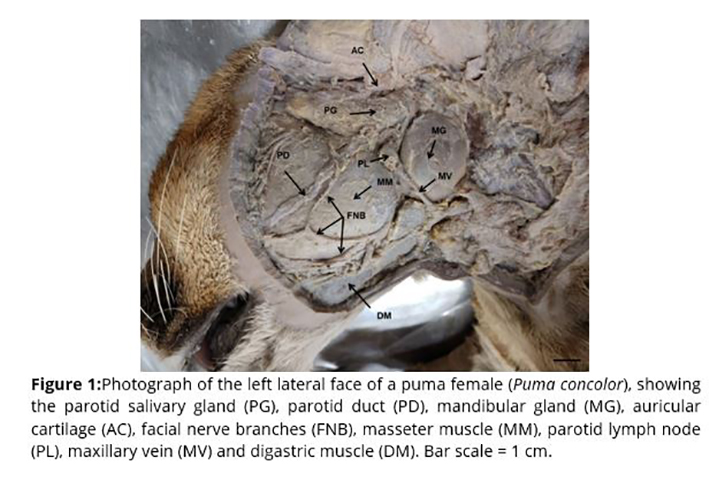
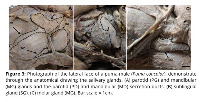
Dissections revealed that the parotid, mandibular, molar and sublingual salivary glands of the puma are distributed throughout the animal's facial region and are drained by rectilinear ducts that converge to the terminal secretory portions. Macroscopically, the parotid gland has a yellowish gray coloration, is distinctly lobulated and has a relatively larger volume compared to the mandibular gland. It has an irregularly semilunar shape and a remarkable fissure in its dorsal portion due to its proximity to the base of the auricular pavilion. The parotid gland is located in the dorsal-caudal region of the face and is arranged ventrally in relation to the basal region of the auricular cartilage. At its ventral end, the parotid covers the apparent origin of the facial nerve and has numerous radicles that originate the parotid duct, which is responsible for draining saliva into the oral vestibule. The parotid duct presents a rectilinear course over the lateral face of the masseter muscle, extending parallelly to the buccal branches of the facial nerve. Along its path, the parotid duct forms a depression on the surface of the masseter muscle called the parotid sulcus (Figures 1 and 3A).
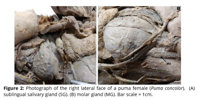
The mandibular salivary gland has a slightly rounded appearance, grayish tint and its surface is lined with a connective tissue capsule. This gland is located in the ventral-caudal region of the face, caudally at the junction angle of the maxillary and lingual-facial veins. Its rostral margin is partially covered by the maxillary vein, while the dorsal-rostral margin is anatomically associated with the parotid lymph node. The mandibular duct originates at the ventral end of the gland, extending deep along the digastric muscle until it reaches the floor of the oral cavity (Figures 1 and 3A). The monostomatic sublingual gland is in direct contact with the rostral edge of the mandibular gland and covered by the mandibular lymph nodes. It has a secretion duct called the larger sublingual duct that usually accompanies the mandibular duct (Figures 2A and 3B). The molar or lingual gland, as it is also known, is a membranous lump of yellowish gray coloration and elongated rectangular shape. Its caudal extremity is dilated and the rostral tapered. This gland is located ventrally to the labial commissure and dorsally to the digastric muscle. Its caudal border is in contact with the masseter muscle and close to the upper labial vein (Figures 2B and 3C).
Discussion
Salivary glands are attachment organs of the digestory system that play an important role in the digestion of food. In addition, its secretions help in the lubrication of food, facilitating chewing and swallowing(14). However, there is little information on the anatomical aspects of these organs in large neotropical wild feline. Morphological knowledge of salivary glands in wild species is fundamental, as it provides basic information for knowledge about species biology. These data canbeused as conservationstrategies, clinicaltreatmentsandpreservationofendangeredspecies.
Based on the dissections, the anatomical attributes of the parotid gland of the puma resemble morphologically in some respects to other wild and domestic carnivorous mammals. The location of the parotid salivary gland in the caudal-dorsal region of the face, ventrally arranged the basal region of the auricular cartilage, is analogous to that found in some wild carnivorous mammals such as crab-eating-fox(7), crab eating raccoon(15) and coati(16). In addition, these authors also found a fissure in the dorsal end of the gland, which is similar to that found in the puma in our study. The origin of the parotid duct at the ventral end of the gland and the rectilinear path over the medial face of the masseter muscle have also been reported in wild carnivorous mammals(7, 15) and domestic carnivores (dog and cat)(17, 18). Some authors also show that in dogs, cats and humans, small accessory parotid glands can be found along the parotid duct(19, 20), a fact that was not observed in this study in the puma. The semilunar shape observed in the parotid gland of the puma also differed from the pattern found in most studiedspecies. In coati, the parotid has a "U" shape, while in the crab eating raccoon,a"Y"(15). In thecrab-eating-fox, dogs and cats, the parotid is irregularly triangular(7, 20).
The morphological and topographic attributes of the puma mandibular gland corroborate numerous aspects with other domestic carnivorous mammals, such as dogs, cats(18, 20) and wild carnivores, such as the undersea, coati(16) and crab-eating-fox(7). However, we observed some discordant anatomical characteristics compared to other wild and domestic species. The medial margin of the mandible gland in the crab-eating-fox, for example, is partially covered by the parotid(7), a feature not found in the puma. The maxillary vein path in dogs runs along the caudal margin of the mandibular gland, diverging from that of the puma in which the vein transits the rostral margin(18). According to Martinelli and Volpi(21), these slight anatomical variations observed in the animals' mandibular gland may be related to small dietary modifications, mainly due to the foods found in different habitats. The anatomical characteristics of the sublingual gland corroborate those found for domestic carnivorous mammals, such as dogs and cats(18, 20) and wild carnivores, such as hand-naked raccoon and coati(16). In addition, the secretion duct of this gland (larger sublingual duct) is similar to that observed for dogs and cats(18, 20) and crab eating raccoon and coati(16). Molar salivary glands are described in the literature only in domestic cats(22, 23), however, a similar shape and location structure has been described in cuicas (Glironia venusta)(24). In dogs, the molar glands are not found(20).
Although there are structural differences, numerous anatomical similarities were observed in the jaguar's salivary glands with the other carnivorous mammals (domestic and wild). These similarities are probably due to the phylogenetic origin of these species. These animals are possibly very close phylogenetically, i.e. derived from the same common ancestor. Thus, there is a high conservative degree of anatomical structures present in these species, which would confirm the hypothesis of a common origin, in which the current forms originated from a possible common ancestor(24).
Conclusion
The dissections revealed that the morphological and topographic aspects of the major salivary glands of the puma follow the same structural pattern described for other carnivorous mammal species (domestic and wild).
References