DOI:
10.1590/1809-6891v20e-5408
MEDICINA VETERINÁRIA
MONITORING OF PROGESTERONE AND ESTRONE FECAL METABOLITES
THROUGHOUT GESTATION IN EWES
AVALIAÇÃO DOS METABÓLITOS FECAIS DE PROGESTERONA E ESTRONA EM OVELHAS
DURANTE A GESTAÇÃO
Rodrigo de Souza Amaral1* ORCID http://orcid.org/0000-0002-0455-2481
Mayara Fonseca Ferreira1 ORCID http://orcid.org/0000-0001-7240-7895
Barbara Luiza Migueis Nunes2 ORCID http://orcid.org/0000-0002-8387-5333
Lais Almeida Gomes1 ORCID http://orcid.org/0000-0002-5992-5839
Arthur Nascimento de Melo3 ORCID http://orcid.org/0000-0002-6261-7730
1Instituto Federal de Educação, Ciência e
Tecnologia do Amazonas, Manaus, AM, Brazil.
2Universidade Federal de Pelotas. Pelotas, RS, Brazil.
3Universidade Federal de Sergipe, Nossa Senhora da Glória, SE,
Brazil.
*Autor para correspondência – rodrigo.amaral@ifam.edu.br
Received on July 28th, 2018.
Accepted on June 17th, 2019.
Introduction
Sheep farming is one of the main livestock, being increasingly developed in all regions of Brazil(1). Thus, the need to improve management protocols and the use of reproductive biotechnologies is extremely important to increase production quantity and quality(2). Hormonal monitoring is a useful tool to support the development of reproductive biotechnologies, enabling the monitoring of endocrine responses to hormonal protocols and applied biotechnologies.
Sheep are also widely used in research as an experimental model for reproductive studies of wild ruminant artiodactyls and for human neonatology studies(3-9). Thus, hormonal monitoring becomes essential for understanding reproductive physiology and monitoring gestational development.
Progesterone is the hormone responsible for the maintenance of pregnancy. In ewes, progesterone is initially produced only by the corpus luteum, but after approximately 55 days of gestation, the placenta produces significant amounts of progesterone that are able to maintain pregnancy, regardless of the presence of a functional corpus luteum(10). Serum estrone levels are elevated at approximately two days before delivery and are related to the triggering of the mechanisms of delivery(10).
However, the technological progress of sheep production must respect the growing concern for animal welfare, understanding its advantages toward the final product(11). Additionally, the use of noninvasive methods for hormonal monitoring during animal experimentation, rather than blood collection decreases the manipulation of individuals and reduces the deleterious effects of stress.
Considering the metabolism and excretion pathways of reproductive steroid metabolites(12), some researchers have evaluated the use of fecal samples for hormone monitoring in sheep. Fecal metabolites of progesterone were evaluated during the estrous cycle and pregnancy in domestic (Ovis aries)(13, 14) and wild (Ovis canadensis)(5) ewes, observing a high correlation between fecal metabolites and serum progesterone levels. Additionally, Schoenecker et al.(6) also evaluated progesterone and estrone fecal metabolite levels in pregnant ewes, but the hormone levels were not correlated with the serum levels.
According to Palme(15), when using alternative biological matrices, such as feces, it is essential to perform physiological validation, demonstrating that the technique used is able to detect changes in the levels of fecal steroid metabolites related to the respective changes in the serum concentrations of these steroids.
Thus, the aim of this study was to monitor progesterone and estrone fecal metabolite levels throughout gestation in ewes, correlating these factors with the serum levels of these steroid hormones.
Material e methods
We used five healthy and cyclic ewes, all housed in the livestock sector of the Federal Institute of Education, Science and Technology of the Amazonas (IFAM), campus Manaus Zona Leste – CMZL, Manaus - AM, Brazil (3.081899S; 59.936908W). Blood and fecal samples were collected between April and October. Extensive daily management was adopted, and the animals were maintained on a pasture during the day and housed at night, with ad libitum water supply and mineral salt.
Initially, the females were monitored for three weeks. During this premating phase, fecal samples from each female were collected twice a week shortly after defecation or taken from the rectal ampoule, and blood samples were collected by venipuncture of the jugular vein once a week. Then, the females were maintained full time with a ram and monitored daily to record the mating, adopting a natural breeding system. The pregnancies were confirmed by ultrasound at 25 days after coverage, and the deliveries were assisted.
After pregnancy confirmation, fecal samples were continuously collected twice a week for up to two weeks after delivery. Blood samples were collected every 15 days during the gestation and postpartum phases. The blood samples were centrifuged for serum separation and stored together with the fecal samples at -20°C until analysis. For adverse reasons, the blood and feces samples of from an ewe were collected only until the 95-day of pregnancy.
All experimental procedures were approved by the IFAM Animal Ethics in Research Committee (Protocol: CEUA.008.02.1928.2206/2017). Fecal samples were lyophilized and subsequently submitted to hormone extraction using 80% methanol, following the protocol described by Palme(15). Briefly, 0.2 g of dried feces was weighed and transferred to a glass tube containing 5 mL of 80% methanol. The tube was shaken for 16 h and then centrifuged. The supernatant (fecal extract) was transferred to a plastic tube and kept at -20°C. An aliquot of the fecal extract was evaporated and then resuspended in buffer (0.04 M NaH2PO4.H2O; 0.06 M Na2HPO4; 0.15 M NaCl; 1.0% BSA; pH 7.0) in the same volume.
For hormone extraction from serum samples, the protocol using diethyl ether as described by Rasmussen et al.(16) was used, followed by resuspension in buffer solution. Samples extracted from serum and feces were analyzed by enzyme immunoassay using a protocol described for several other species(17-19). Antibodies CL425 (1: 5,000) for progesterone and R522-2 (1: 20,000) for total estrone, with their respective peroxidase-conjugated hormones (1: 160,000; 1: 350,000), all provided by the University of Davis – UC Davis, USA, were used.
Microtiter plates (MaxiSorp, Nunc, Rochester, USA) were coated (50 μL/well) with antibody diluted in labeling solution (0.015 M Na2CO3.H2O, 0.035 M NaHCO3; pH 9.6), sealed with acetate adhesive and incubated at 4°C for 16 h. After incubation, the plates were washed three times (0.15 M NaCl, 0.05% Tween-20). Then, 25 μL of buffer solution was added to each well, and 50 μL of each sample, standard or control was added. Then, 50 μL of enzyme-conjugated hormone solution diluted in buffer solution was immediately added. The plates were sealed and incubated for 2 h at room temperature.
After incubation, the plates were washed and then 100 μL/well of substrate solution (250 µL of 0.016 M tetramethylbenzidine in dimethylsufoxide; 50 µL of 0.1752 M H2O2; 11 mL of substrate buffer [0.01 M C2H3Na; pH 5.0]) was added. The chromogenic reaction was stopped with 50 μL of acidic solution (4.0 M H2SO4). The optical density of each well was measured on a plate reader using a 450 nm filter.
All samples, controls and standards were analyzed in duplicate. The sensitivities of the progesterone and estrone assays were 0.07 ng/mL and 0.08 ng/mL, respectively. The intra- and interassay coefficients of variation of the high (70% binding) and low (30% binding) controls were <10.35% for both assays, and all assays showed parallelism between serial dilutions of the samples and the standard curve of the assay. Serum progesterone levels were presented as ng/mL and estrone levels were presented as pg/mL, whereas progesterone and estrone fecal metabolite results were corrected and presented as ng/g of dry feces.
For data standardization, only hormonal results from samples between 14 days before mating and 15 days after delivery were used. Hormone data were aligned considering the day of mating and the hormone profile plotted. The correlation between serum hormone levels and their fecal metabolites was calculated (Pearson’s correlation). Hormone levels were also separated into three thirds of pregnancy and statistically compared (Kruskal-Wallis test and Tukey’s post hoc test; Bioestat Program, IDSM). A probability value of P < 0.05 was considered significant.
Results
The average duration of gestation was 147.2 ± 6.5 days, ranging from 143 to 157 days, totaling approximately 21 weeks. One of the five ewes showed a twin pregnancy.
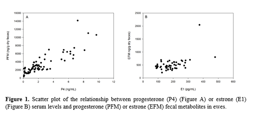
Serum and fecal hormone profiles showed a significant positive correlation (R = 0.8572, P < 0.001 for progesterone and R = 0.5893, P < 0.001 for estrone) (Figure 1).
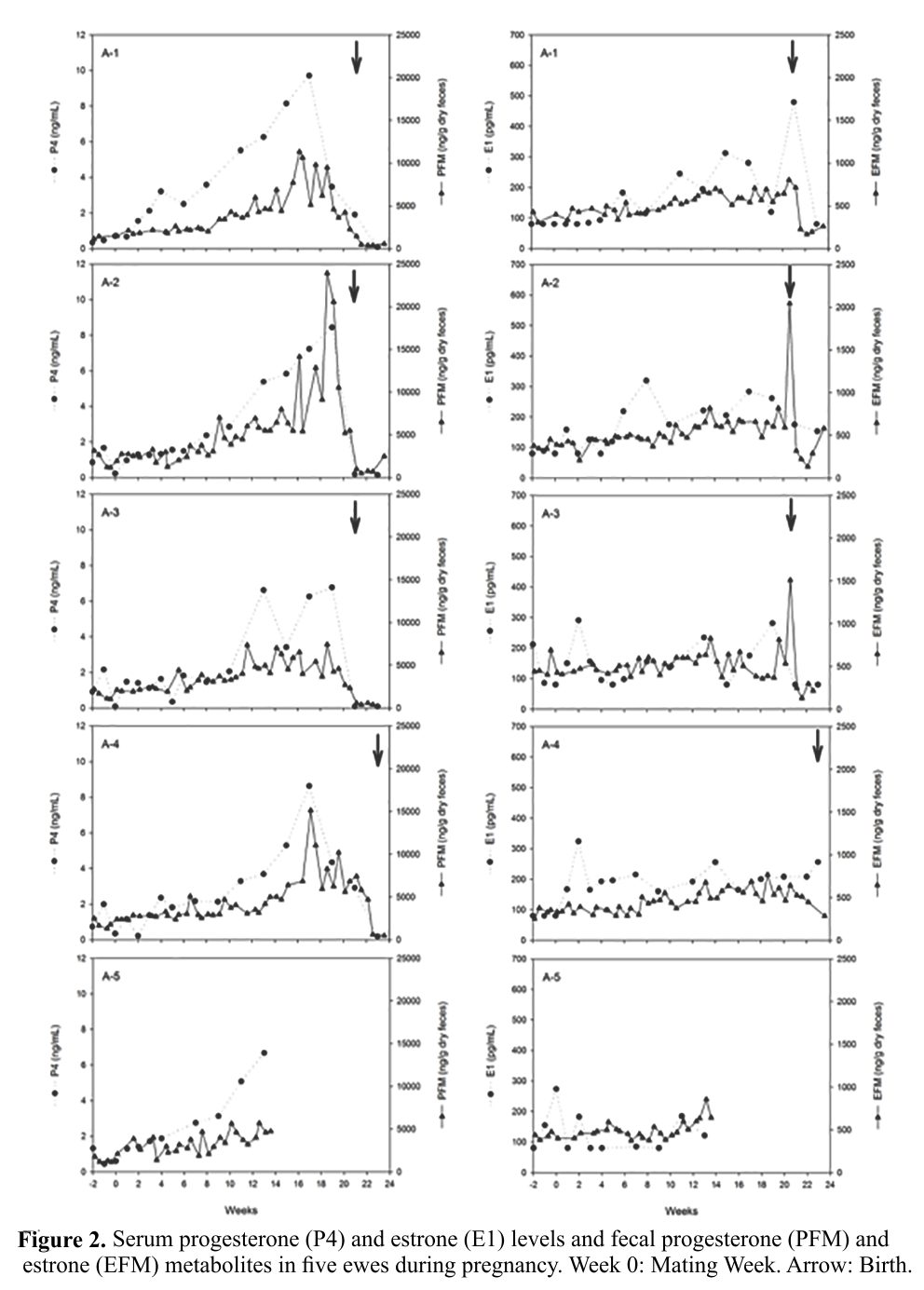
The female with the twin pregnancy (Figure 2, A-1) did not show discrepant hormone values from the other females. For this female, the serum progesterone levels and their fecal metabolites during pregnancy ranged from 0.7 to 9.7 ng/mL and 1,489.0 to 11,324.8 ng/g of dry feces, respectively, while for the remaining females these levels were 0.2 to 8.6 ng/mL and 1,254.2 to 23,931.8 ng/g of dry feces, respectively. For serum estrone and its fecal metabolites, the values for the twin pregnancy female were 80.0 to 479.2 pg/mL and 335.5 to 803.3 ng/g of dry feces, respectively, and for the other females were 80.0 to 323.8 pg/mL and 206.9 to 2.044.9 ng/g of dry feces, respectively.
After mating, all females showed an increasing profile of progesterone serum levels during pregnancy, with a subsequent decline to basal levels after parturition (Figure 2). The same profile was observed for progesterone fecal metabolite levels (Figure 2). Serum estrone levels showed a profile with a slight increase during pregnancy, showing higher values on the week of delivery, wherein the hormone peak on the week of delivery was more evident for fecal samples (Figure 2).
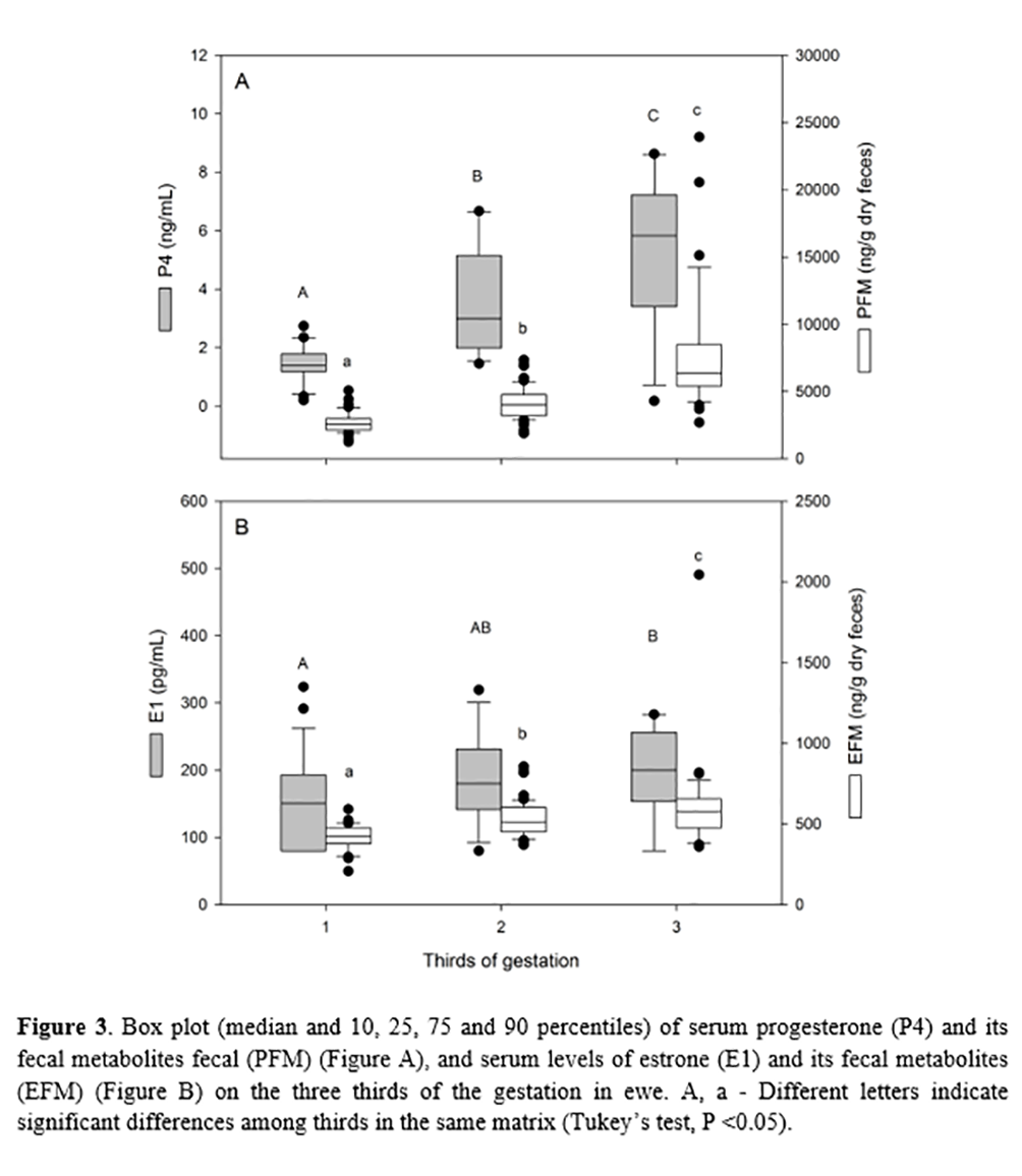
Comparing hormone levels among three-thirds of gestation in single pregnancy animals, progesterone levels showed significantly increasing values in both matrices (P < 0.001, Tukey’s test, Figure 3). For estrone, fecal metabolite levels of estrone also showed increasing values, with significantly higher levels in the final third of pregnancy than the initial and middle thirds (P < 0.010, Tukey test). The rise in serum estrone levels during pregnancy, however, was slight, with values for the final third being statistically higher than the initial third (P < 0.050, Tukey’s test, Figure 3).
Discussion
The mean pregnancy length on the monitored ewe and the observed individual variation corroborate those reported in the literature for the species(20). The blood hormone profile during pregnancy in ewes has been previously described(21-23), and the serum progesterone and estrone levels during pregnancy obtained in this study corroborated previous reports. The high correlation between serum progesterone levels and fecal metabolites during pregnancy was also observed by Cebulj-Kadunc et al.(14) in ewes. Additionally, Borjesson et al.(5) also demonstrated a high correlation between serum and fecal progesterone levels in wild ewes. In goats, Capezzuto et al.(24) also observed a high correlation between progesterone and estradiol serum levels and their fecal metabolites. Thus, the results of the present study reinforce the advantage of using fecal rather than serum samples for longitudinal progesterone monitoring throughout gestation in ewe, minimizing the stress of handling animals.
However, although Schoenecker et al.(6) previously evaluated fecal metabolite levels of estrone during pregnancy in two wild ewes, there is no report of the correlation level between serum estrone levels and their fecal metabolites. Thus, the present study demonstrates the existence of a significant correlation of estrone between the two biological matrices evaluated during pregnancy.
According to Palme et al.(12), the main route of progesterone and estrone excretion in sheep is fecal, in which approximately 76% of progesterone metabolites and 88% of estrone metabolites are excreted in feces. Additionally, according to these authors, the metabolism and excretion of these hormones in feces occur in less than 24 h. The rapid metabolism of progesterone and estrone and preferential fecal excretion justifies the correlation observed between these hormones and their fecal metabolites.
Both biological matrices showed a significant increase in the progesterone profile during pregnancy, with a subsequent decline to baseline levels, corroborating the literature, as expected. According to Noakes et al.(10), ewes with approximately 55 days of gestation (~ 7 weeks) show a gradual increase in serum progesterone levels, related to the onset of the production of this hormone by the placenta.
Considering the endocrine role of the placenta in progesterone production to pregnancy maintenance, according to Bassett et al.(25), ewes with twin pregnancies may have up to twice the blood levels of progesterone during the final third of pregnancy compared to ewes with single pregnancy. In the present study, only one female had twin pregnancies, which did not show discrepant hormone values from those of females with a single pregnancy. The metabolism and excretion rate of fecal progesterone metabolites may vary among individuals of the same species(12). Therefore, it is possible that the lack of marked variations in the levels of fecal metabolites of progesterone between the twin pregnant female and the other single pregnant females may be related to variations in the individual metabolism rate. Therefore, further study comparing animals from different gestational conditions is recommended to better understand fecal progesterone metabolite levels in multiple pregnancies in sheep.
For estrone, according to Tsang(22), Thompson and Wagner(26) and Noakes et al.(10), its serum levels in pregnant ewes are greatly increased within two days before calving, being related to the induction parturition mechanisms. This information corroborates the statistically higher values of estrone in the final third of pregnancy for both matrices observed in this study. Additionally, it was possible to observe an increase in fecal metabolite levels of estrone in the week of delivery of the monitored animals.
However, despite the results obtained in the fecal matrix corroborating the estrone profile reported in the literature, this result was not clearly observed in serum estrone levels. This discrepancy is related to the low frequency of blood collection used in this study during pregnancy (every 15 days), which did not alloe the sampling of serum estrone peak levels in all females. Capezzuto et al.(24) also observed a better estrogen hormone pattern in fecal samples than in serum samples when evaluating weekly goat samples during pregnancy. Considering the entire process of metabolization and the excretion of hormones involved in the present study, as already described by Palme et al.(12), the possibility of monitoring specific physiological variations, such as peripartum elevation of estrone levels, using fecal samples is demonstrated, thus reducing the intensity in the handling of animals.
The increasing use of sheep as an experimental model in reproductive studies of wild ruminant Artiodactyls and in human neonatology studies(3-9) makes the development of noninvasive hormonal monitoring techniques, such as the use of fecal samples, an important support tool for these studies. Thus, the results obtained in the present study demonstrate the possibility of endocrine monitoring of pregnant ewes submitted to experimental neonatology protocols, minimizing the deleterious effects of stress and highlighting this analysis as a tool to be used in wild sheep and tested in wild deer.
Conclusion
This study showed that changes in progesterone and estrone fecal metabolite levels during pregnancy reflect the expected physiological serum changes of these hormones in ewes, demonstrating the feasibility of noninvasive longitudinal monitoring of progesterone and estrone levels using fecal samples in this species.
Acknowledgments
We thank the Coordenação Geral de Produção (CGP/IFAM-CMZL) for the support on the management of animals. We also thank PR-PPGI/IFAM for the fellowship (MFF and BLMN: IC-IFAM; RSA: Productivity/IFAM). This project was financed by a PADCIT/PR-PPGI/IFAM-2016 grant.
References
1.
Martins
EC, Magalhães KA, Souza JDF, Guimarães VP, Barbosa CMP, Holanda Filho
ZF. Cenário mundial e nacional da caprinocultura e da ovinocultura.
Boletim ativos de ovinos e caprinos. 2016;3(2):3-6.
2. Simplício AA, Freitas VJF,
Fonseca JF. Biotécnicas da reprodução como técnicas de manejo
reprodutivo em ovinos. Revista Brasileira de Reprodução Animal.
2007;31(2):234-46.
3. Oishi PE, Sharma S, Datar SA, Kumar S, Aggarwal S, Lu Q, et al. Rosiglitazone preserves pulmonary vascular function in
lambs with increased pulmonary blood flow. Pediatric Research.
2013;73(1):54-61.
4. Smolich JJ, Mynard JP. Increased right ventricular power and ductal
characteristic impedance underpin higher pulmonary arterial blood flow
after betamethasone therapy in fetal lambs. Pediatric Research.
2018;83(7):1-6.
5. Borjesson DL, Boyce WM, Gardner IA, DeForge J, Lasley B. Pregnancy
detection in bighorn sheep (Ovis
canadensis) using a fecal-based enzyme immunoassay. Journal of
Wildlife Diseases. 1996;32(1):67-74.
6. Schoenecker KA, Lyda RO, Kirkpatrick J. Comparison of three fecal
steroid metabolites for pregnancy detection used with single sampling in
bighorn sheep (Ovis canadensis).
Journal of Wildlife Diseases. 2004;40(2):273-81.
7. Jabbour HN, Hayssen V, Bruford MW. Conservation of deer:
contributions from molecular biology, evolutionary ecology, and
reproductive physiology. Journal of Zoology. 1997;243(3):461-84.
8. Andrabi SMH, Maxwell WMC. A review on reproductive biotechnologies
for conservation of endangered mammalian species. Animal Reproduction
Science. 2007;99(3-4):223-43.
9. Thongphakdee A, Berg DK, Tharasanit T, Thongtip N, Tipkantha W,
Punkong C, et al. The impact of ovarian stimulation protocol on oocyte
quality, subsequent in vitro embryo development, and pregnancy after
transfer to recipients in Eld's deer (Rucervus
eldii thamin). Theriogenology. 2017;91(1):134-44.
10. Noakes DE, Parkinson TJ, England GCW. Veterinary Reproduction and
Obstetrics. 9 ed. London:
Saunders Elsevier; 2009. 950 p.
11. Rufino LAL, Araújo AA. Indicadores de bem estar em ovinos e
caprinos: uma revisão. Revista Brasileira de Higiene e Sanidade Animal.
2015;9(2):294-8.
12. Palme R, Fischer P, Schildorfer H, Ismail MN. Excretion of infused
14C-steroid hormones via faeces and urine in domestic livestock. Animal Reproduction Science.
1996;43(1):43-63.
13. Furtado PV. Perfil analítico de estrógenos e progestinas em
diferentes matrizes biológicas na espécie ovina (Ovis
aries) [Tese]. São Paulo: Universidade de São Paulo; 2007.
doi:10.11606/T.10.2007.tde-11042008-092435
14. Cebulj-Kadunc N, Snoj T, Cestnik V. Faecal gestagen, serum and milk
progesterone concentrations in ewes of the Jezersko-Solchava breed. Acta Veterinaria Brno. 2000;69(1):33-7.
15. Palme R. Measuring fecal steroids: Guidelines for practical
application. Annals of the New York Academy of Sciences.
2005;1046(1):75-80.
16. Rasmussen FE, Wiltbank MC, Christensen JO, Grummer RR. Effects of
fenprostalene and estradiol-17 beta benzoate on parturition and retained
placenta in dairy cows and heifers. Journal of Dairy Science.
1996;79(2):227-34.
17. Munro CJ, Stabenfeldt GH, Cragun JR, Addiego LA, Overstreet JW,
Lasley BL. Relationship of serum estradiol and progesterone
concentrations to the excretion profiles of their major urinary
metabolites as measured by enzyme immunoassay and radioimmunoassay.
Clinical Chemistry. 1991;37(6):838-44.
18. Graham LH, Schwarzenberger F, Möstl E, Galama W, Savage A. A
versatile enzyme immunoassay for the determination of progestogens in
feces and serum. Zoo
Biology. 2001;20(3):227-36.
19. Amaral RS, Da Silva VMF, Lazzarini SM, D'Affonseca Neto JA, Ribeiro
D, Rosas FCW. Assessment of sexual maturity
in captive Amazonian manatees (Trichechus
inunguis). Marine Mammal Science. 2018;34(1):190-9.
20. Hafez ESE, Hafez B. Reproduction in Farm Animals. 7 ed.
Philadelphia: Lippincott Williams & Wilkins; 2000. 509
p.
21. Ranilla MJ, Sulon J, Carro MD, Mantecón AR, Beckers JF. Plasmatic profiles of pregnancy-associated glycoprotein and
progesterone levels during gestation in Churra and Merino sheep.
Theriogenology. 1994;42(1):537-45.
22. Tsang CPW. Plasma levels of estrone sulfate, free estrogens and
progesterone in the pregnant ewe throughout gestation. Theriogenology.
1978;10(1):97-110.
23. Mukasa-Mugerwa E, Viviani P. Progesterone concentrations in
peripheral plasma of Menz sheep during gestation and parturition. Small
Ruminant Research. 1992;8(1):47-53.
24. Capezzuto A, Chelini MOM, Felippe ECG, Oliveira CA. Correlation
between serum and fecal concentrations of reproductive steroids
throughout gestation in goats. Animal Reproduction Science.
2008;103(1-2):78-86.
25. Bassett JM, Oxborrow TJ, Smith ID, Thorburn GD. The concentration of
progesterone in the peripheral plasma of the pregnant ewe. Journal of
Endocrinology. 1969;45(3):449-57.
26. Thompson FN, Wagner WC. Plasma progesterone and oestrogens in sheep
during late pregnancy: contribution of the maternal adrenal and ovary.
Journal of Reproduction and Fertility. 1974;41(1):57-66.