DOI:10.1590/1809-6891v20e-53529
>MEDICINA VETERINÁRIA
OCCURRENCE OF FILARID PARASITES IN HOUSEHOLD AND SHELTERED
DOGS IN THE CITY OF JOINVILLE – SANTA CATARINA, BRAZIL
OCORRÊNCIA DE FILARÍDEOS PARASITOS EM CÃES DOMICILIADOS E PROVENIENTES
DE ABRIGO ANIMAL DE JOINVILLE - SANTA CATARINA, BRASIL
Anna Karina Kannenberg1 ORCID http://orcid.org/0000-0001-6501-2369
Luca
Frondana1 ORCID http://orcid.org/0000-0003-4468-1648
Igor
Henrique Rodrigues Martins1 ORCID http://orcid.org/0000-0003-1355-3686
Charlene
Edianez Longhi1 ORCID http://orcid.org/0000-0003-4515-2756
Michele Müller Fialkowski2 ORCID http://orcid.org/0000-0001-9067-169X
Viviane
Milczewski1* ORCID http://orcid.org/0000-0002-7117-5380
1Federal
Institute of Santa Catarina, Araquari, SC, Brazil.
2Badanievet
Laboratory, Araquari, SC, Brazil.
*Corresponding
author - viviane.milczewski@ifc.edu.br
Abstract
Parasites from the Filariidae family are among the nematodes that can
infest dogs. Some may be apathogenic, such as Acanthocheilonema
reconditum, and others such as Dirofilaria
immitis are potentially zoonotic and may lead dogs to death. Thus,
the aim of this study was to perform a survey on filarid infection in
dogs with a fixed domicile and wandering dogs temporarily sheltered in
the city of Joinville, SC, Brazil. Blood samples from 429 animals were
collected from 2015 to 2017 and submitted to screening for microfilariae
and/or D. immitis antigens.
Our results revealed that twenty-four samples (5.6%) displayed A.
reconditum microfilariae and three samples (0.7%) were positive
for D. immitis, totaling 27
(6.3%) positive animals for filarid parasites. Additionally, A.
reconditum was more prevalent in dogs from shelters, whereas the
three D. immitis positive
samples were from household dogs that displayed no clinical signs of the
presence of parasites. The occurrence of these parasites in dogs from
Joinville indicates the importance of differential diagnosis of
microfilaria species for the correct clinical management of infected
dogs. Our epidemiological information may help the medical and
veterinary community of the region regarding clinical suspicion of
canine and human heartworm disease, as well as guide preventive actions
targeting public and animal health.
Keywords:
Acanthocheilonema reconditum, Dirofilaria immitis,
microfilariae, zoonosis.
Resumo
Dentre
os
diversos nematódeos filarídeos que parasitam cães, alguns podem ser
apatogênicos como Acanthocheilonema
reconditum e outros como Dirofilaria
immitis podem levar esses animais a óbito, além de terem potencial
zoonótico. Nesse sentido, o objetivo deste trabalho foi realizar um
levantamento da ocorrência de filarídeos em cães residentes em domicílio
fixo e cães errantes que estavam
temporariamente em um abrigo animal de Joinville-SC. Entre 2015 e 2017,
amostras de sangue de 429 animais foram submetidas à busca de
microfilárias ou antígenos de D.
immitis, utilizando-se Método de Knott modificado e kit comercial
de diagnóstico clínico. Vinte e quatro amostras (5,6%) apresentaram
microfilárias de A. reconditum
e três (0,7%) foram positivas para D.
immitis, totalizando 27 (6,3%) cães positivos para a presença de
filarídeos parasitos. A.
reconditum teve maior ocorrência em cães provenientes de abrigos,
já os três cães positivos para D.
immitis eram domiciliados e não apresentavam suspeita clínica de
dirofilariose. A presença desses parasitos em cães da região denota a
importância do diagnóstico diferencial das espécies de microfilárias
para o encaminhamento clínico adequado dos cães parasitados. As
informações epidemiológicas obtidas podem orientar a comunidade médica e
veterinária em relação à atenção para as suspeitas clínicas de
dirofilariose canina e humana, assim como na orientação das medidas de
prevenção visando a saúde pública e animal.
Palavras-chave:
Acanthocheilonema reconditum, Dirofilaria immitis,
microfilárias, zoonose.
Received on June 15, 2018
Accepted on April 25, 2019
Introduction
Filarids (Filarioidea superfamily) are nematode parasites that are located in different organs of the definitive host and present a biological cycle characterized by the release of microfilariae in the circulation, which are ingested and transmitted by a hematophagous intermediate host.
In Brazil, several genera of filarids have been reported in domestic and wild mammals. Moraes et al.(1) found Acanthocheilonema reconditum, Brugia sp., Dirofilaria immitis, Dirofilaria (Nochtiella) repens, and Mansonella sp. in coatis (Nasua nasua) in a preservation area of the Atlantic forest in Foz do Iguaçu, Paraná.
In the canine species, there has always been great interest in the identification of filarid parasites due to clinical relevance or possible zoonotic potential. Several filariae of domestic dogs have been reported around the world, such as Brugia pahangi(2), Dirofilaria (Nochtiella) repens(3), Dipetalonema dracunculoides(4), Brugia malayi(5), and Cercopithifilaria bainae(6). The latter was recently reported in dogs in the Northeast region of Brazil(7).
As in other regions of the world, Dirofilaria immitis and Acanthocheilonema reconditum are the most prevalent filarids in Brazil(7–10). Filariae of Acanthocheilonema sp. are transmitted biologically by fleas (Ctenocephalides canis, Ctenocephalides felis, Pulex irritans, P. simulans, and Echidnophaga gallinae) or hematophagous lice (Heterodoxus spiniger and Linognathus setosus) during blood-sucking(11). They are considered poorly pathogenic probably because they preferentially parasitize the subcutaneous tissue(12). On the other hand, D. immitis is transmitted by several mosquitoes, such as Aedes vexans (Meigen), Anopheles quadrimaculatus (Say), Anopheles punctipennis (Say), Culex pipiens quinquefasciatus (Say), Culex erraticus (Dyer and Knab), Culiseta inornata (Williston), Psorophora columbiae (Dyer and Knab), Psorophora ferox (Humboldt), and Psorophora howardii (Coquillett)(13). D. immitis parasitize preferentially domestic and wild canids. At the adult stage, it is installed in the pulmonary artery and right ventricle and may cause signs of apathy, exercise intolerance, circulatory disorders such as vena cava syndrome or death.
Brazil presents a wide variation in the prevalence of heartworm disease among its different regions(10). According to Labarthe and Guerrero(14), coastal regions have a higher occurrence of heartworm disease due to the reproductive cycles of vector mosquitoes. The city of Joinville, located in northern Santa Catarina, bordered by the Babitonga Bay, and counting with 36 km2 of mangrove area, has climatic characteristics propitious for the spread of vectors(15). It presents mean monthly precipitation of 183.6 mm(16) and annual means of temperature and humidity of 22.4 °C and 80.76%, respectively(17).
Although Bublitz et al.(18) have reported eight cases of human heartworm disease in Joinville, there are no studies that indicate the prevalence of dogs parasitized by filariae in the city. Thus, this study aimed to perform a survey on the occurrence of filarid parasites of household dogs and dogs from animal shelters in the city of Joinville, SC, Brazil.
Material and methods
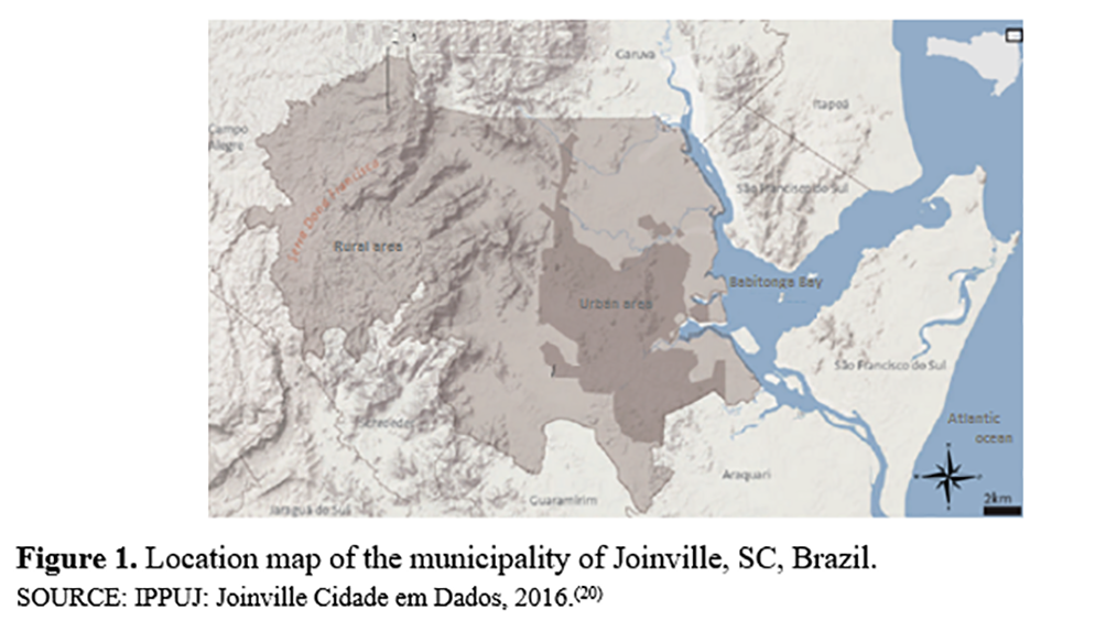
This study was carried out in the city of Joinville, located in the northern coast of the state of Santa Catarina (26°18′14″ S and 48°50′45″ W), in the South of Brazil. It has a total extension of 1,124.10 km2, with 913.7 km2 of a rural area and 210.4 km2 of an urban area(15), bordering the Serra do Mar and Babitonga Bay (Figure 1). The altitude ranges from zero to 1,325 meters and, according to the classification of Köppen and Geiger(19), the regional climate is Cfa, described as humid to super humid, dependent on the Atlantic Polar mass (winter and autumn) and Atlantic Tropical mass (summer and spring) for the season and temperature changes. There is no dry season, and temperatures range from 31 °C (maximum means) in January to 13 °C (minimum means) in July, with mean annual relative air humidity of 76.04%.
The studies were conducted in three phases from 2015 to 2017, totaling 429 dogs over six months of age, household (considering animals residing in fixed domiciles with responsible guardians) or coming from rescues and temporarily sheltered animal centers of Joinville. None of the dogs participating in the study had a clinical suspicion of heartworm disease.
The first phase of this study was carried out from May to September 2015 using 196 blood samples from household dogs. Samples were collected from dogs treated at veterinary clinics in the city, which had laboratory tests required by clinical veterinarians for various reasons and referred to the clinical laboratory service provider. All the excess material after the tests requested by the clinician were sent to the research laboratory and used in our study.
The second phase comprised the period from October 2015 to October 2016, with 95 blood samples from dogs sheltered by the Animal Shelter of the city of Joinville, identified by sex and animal stall number.
The third phase, carried out between May and December 2017, involved 138 blood samples from dogs domiciled in the city of Joinville, taken by their guardians to veterinary consultations for several reasons. Data on sex and the possible presence of clinical signs indicative of heartworm diseases, such as anorexia, weight loss, apathy, cough, and exercise intolerance, were collected. The study included dogs who have not received heartworm disease preventive drugs within 30 days or microencapsulated moxidectin (ProHeart® SR-12) within one year before sampling collection. The research project was approved by the Ethics Committee of the Federal Institute of Santa Catarina, Araquari Campus, under the number 0177/2016.
The samples were stored under refrigeration in tubes with EDTA and sent to the Laboratory of Veterinary Teaching and Diagnosis – Parasitology of the Federal Institute of Santa Catarina, Araquari Campus, for processing. The whole blood was subjected to the modified Knott technique(21), and the plasma obtained by centrifugation was frozen at −18 °C. The twenty-four microfilaremic samples found throughout the experiment had larvae measured under an Olympus microscope using the Software Cellsens Standard® program. All samples of the second and third phases, as well as those that presented microfilariae in the first experiment, were submitted to the SNAP 4Dx® Plus clinical diagnostic kit, IDEXX. Additionally, samples from the first and second phases were also evaluated in the thick-drop method(22).
The frequencies of D. immitis and A. reconditum infection according to sex and origin of dogs (household and sheltered) were compared using the chi-square test, rejecting the null hypothesis at 5% significance level. The program PAST v. 3.16(23) was used. The continuity-correction chi-square test (McNemar’s test) was used to compare the sensitivity of microfilariae detection methods.
Results and discussion
Considering the canine population used in this study (n=429), 6.3% presented parasitism by filarid, 5.6% positive for A. reconditum and 0.7% for D. immitis. In prevalence studies in coastal cities located from 74 km north of Joinville, Leite et al.(24) found in the city of Guaratuba, PR, and Reifur et al.(9) on the coast of Paraná 0.94% and 5.47% of dogs positive for D. immitis, respectively, and 22.6% positive for A. reconditum. Similarly, in coastal cities located 80 km south of Joinville, Lira et al.(25) verified 4.8% of dogs parasitized by A. reconditum and none by D. immitis in Itajaí, SC, whereas Araújo et al.(26) found 15% of dogs positive for D. immitis and none for A. reconditum in Florianópolis, SC. Variations of occurrences of filarids in different geographical areas are correlated to the availability of definitive hosts, the possibility of being parasitized by intermediate hosts, and environmental conditions that lead to the multiplication of populations of these hematophagous arthropods. Joinville has environmental conditions that promote the maintenance of multiplication of transmitting mosquitoes. According to Silva and Langoni(27), the frequency of bloodsucking in the canine population is constant in areas where conditions are favorable to the presence of infected mosquitoes throughout the year and may increase the agent transmission rate. In addition, the municipality has the Dona Francisca Environmental Preservation Area, where 112 species of wild mammals, including procyonids and canids as potential hosts of D. immitis, were registered(28). Regarding the transmission of D. reconditum, carried out by fleas, parasite dispersion may be influenced by specific conditions of the microenvironment where the dog remains. In our study, sheltered dogs had a higher prevalence of A. reconditum (p=1.99×10⁻⁸; c2=31.50) in relation to household dogs (Table 1).

Animal confinement in large groups and difficulties in controlling fleas in shelter environments may have contributed to the transmission of A. reconditum. Regarding sex, a higher prevalence of A. reconditum was observed in males among household dogs (p=0.01; c2=6.38), with no significant difference between sex for sheltered dogs (p=0.21; c2=1.56). Brito et al.(29) in Brazil and Brianti et al.(11) in Italy also observed no significant differences in the occurrence of this parasite between males and females. However, Hashem and Badawy(30) found a higher prevalence in males. These authors related the susceptibility of these dogs to the infection to possible hormonal effects. In our study, sheltered dogs may not have presented a difference of prevalence according to sex because 68 animals were castrated (70%), thus preventing the hormonal manifestation of susceptibility. Two of the dogs with A. reconditum had clinical signs reported by veterinarians, such as joint pain, lethargy, inappetence, anorexia, weight loss, and cough. Although Hashem and Badawy(30) stated that hemolytic anemia with impaired hepatic and renal function are the main results of A. reconditum infection, it is not possible to state that the alterations found in this study have relations with the parasite since the animals could have other diseases concomitant with parasitism.
Microfilariae were found in both used parasitological methods, but the modified Knott method (Newton and Wright, 1956) was more sensitive (p=0.003; c2=9.09) (Table 2) when compared to the thick-drop method, in addition to allowing the morphological evaluation of microfilariae, favoring species differentiation. According to Dillon et al.(31), the animal must have at least 20 to 50 microfilariae/mL of blood to be detected by the thick-drop method.

The 4Dx® Plus SNAP rapid diagnostic test detects circulating D. immitis antigens and has, on average, 97.5% sensitivity and 94% specificity(32). In our study, the three dogs seropositive for D. immitis presented detectable antigens by this diagnostic method, but no microfilariae were found in one of these samples. Occult heartworm disease can occur due to the presence of single-sex parasites, low infection rate(33), microfilaricidal drug-treated animals, and in dogs that have developed an efficient immune response to microfilariae(8). Antigenemia, caused by adult females, usually precedes the appearance of microfilariae in the blood(34). Initially, the three infected dogs detected in this study were evaluated by clinical veterinarians and had no clinical suspicion of heartworm disease, although one had cough and the other apathy, which could be compatible with parasitism. A blood count was requested to assist the diagnosis of the cases, and microfilariae were found at the time of blood smears. Then, these samples were processed by the team of this experiment, when the presence of microfilariae compatible with D. immitis and positivity in the antigenic test was confirmed. The third dog was suspected of having ehrlichiosis and had antibodies against Ehrlichia canis or Ehrlichia ewingii and D. immitis when the blood was submitted to SNAP 4Dx® Plus. Subsequently, this sample was processed by our team, confirming the presence of D. immitis antigens and absence of microfilariae in the circulation. The absence of clinical signs can often be observed in parasitized dogs. Larsson et al.(34) found that 42.86% of dogs infected with D. immitis did not present changes in the clinical evaluation. Similarly, Meriem-Hind and Mohamed(35) found that 75.56% of the dogs were asymptomatic carriers. The absence of signs is related to the number of parasites present, the individual immune response, and the level of exercises to which the dog is submitted(36).
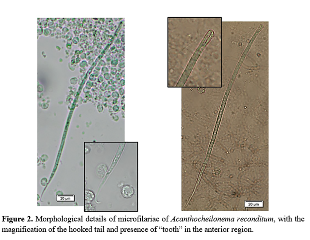
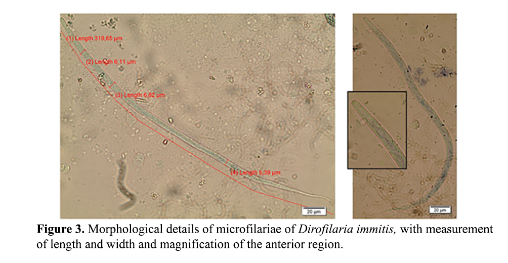
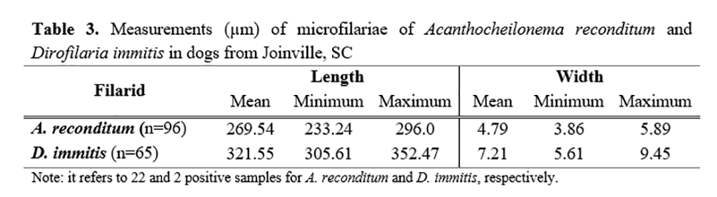
Regarding the morphology of microfilariae, positive samples for A. reconditum had larvae of typical appearance(7), with rounded anterior and presence of “tooth,” and some of them with a hooked tail (Figure 2; Table 3). On the other hand, microfilariae of D. immitis were larger, with conical anterior portion and straight tails (Figure 3; Table 3).
All positive samples for microfilariae of A. reconditum were negative for the presence of D. immitis antigens. The mean length of microfilariae of A. reconditum (269.54 µm) (Table 3) was similar to that found in dogs from the Northeast of Brazil (272.3 ± 4.3 µm)(7) and Italy (273.1 ± 9.4 µm)(11).
However, when considering the minimum and maximum measurements, we observed 19.8% of microfilariae of A. reconditum out of the limit dimensions of 250–288 µm in length, as limits suggested by Zajac and Conboy(37). Microfilariae of D. immitis presented a mean of 321.55 µm in length, which is similar to those found by Reifur et al.(9) (323.49 ± 12.73 µm), who studied dogs from the coast adjacent to those of our experiment. However, Ramos et al.(7) found smaller measurements in the state of Pernambuco (301.2 ± 7.6 µm). Among the 65 microfilariae of D. immitis measured from the two microfilaremic dogs, 19 (29.23%) presented length above the limit suggested by Zajac and Conboy(37). Reifur et al.(9) found specimens of microfilariae with distinct morphology, not identified by the authors. These data indicate that there are metric and morphological variations among microfilariae found in dogs and should be better studied.
Conclusions
This study is the first report of the presence of D. immitis in Joinville, SC. The presence of A. reconditum and D. immitis in native dogs indicates the existence of epidemiological conditions (environment – host – etiological agent) for the transmission and maintenance of these filariae in the region. Considering that A. reconditum is an apathogenic parasite and heartworm disease may be a serious disease, the morphological differentiation of microfilariae in the circulation of dogs from the region is essential. The epidemiological information obtained may help the medical and veterinary community regarding the clinical suspicion of canine and human heartworm disease, as well as guide preventive actions targeting public and animal health in the inhabitants of the region.
Acknowledgments
To Dr. Norma Labarthe and the directors of the Animal Shelter of Joinville. The Research and Innovation Support Foundation of Santa Catarina State (FAPESC) and Federal Institute of Santa Catarina (IFC) funded this project.
References
1.
Moraes MFD; Silva MX; Matos PCM; Albuquerque ACA; Tebaldi JH; Mathias
LA; Hope EGL. Filarial nematodes with zoonotic potential in ring-tailed
coatis (Nasua nasua Linnaeus, 1766, Carnivora: Procyonidae) and
domestic dogs from Iguaçu National Park, Brazil. Veterinary
Parasitology: Regional Studies and Reports. 2017;8:1-9. Available at: https://doi.org/10.1016/j.vprsr.2017.01.003.
2. Schacher JF; Sahyoun PF. A chronological study of the
histopathology of filarial disease in cats and dogs caused by Brugia
pahangi (Buckley and Edeson, 1956). Transactions of the Royal
Society of Tropical Medicine and Hygiene, 1967. 61(2):234-236. Available
at: https://doi.org/10.1016/0035-9203(67)90162-9
3. Hermosilla C; Pantchev N; Dyachenko V; Gutmann M; Bauer
C. First autochthonous case of canine ocular Dirofilaria
repens infection in Germany – Short Communication. Veterinary
Record, 2006. 158(4):134-135. Available at: http://dx.doi.org/10.1136/vr.158.4.134.
4. Giannetto S; Poglayen G; Gaglio G; Brianti E; Ferlazzo
M; Giudice E. Dipetalonema
dracunculoides (Nematoda: Onchocercidae): first report in dog in
Italy. Parasite, 2003. 10(2):188.
5. Ravindran R; Varghese S; Nair SN; Balan VM; Lakshmanan B; Ashruf RM;
Kumar SS; Gopalan AKK; Nair AS; Malayil A; Chandrasekhar L; Juliet S;
Kopparambil D; Ramachandran R; Kunjupillai R; Kakada SAM. Canine
Filarial Infections in a Human Brugia
malayi Endemic Area of India. Biomed Research International
[Internet]. 2014, May; 2014:1-9. Available from: https://www.hindawi.com/journals/bmri/2014/630160/ https://doi.org/10.1155/2014/630160. English.
6. Ionică A.M.; D'Amico G.; Mitková B.; Kalmár Z.; Annoscia G.; Otranto
D.; Modrý D.; Mihalca A.D. First
report of Cercopithifilaria
spp. in dogs from Eastern Europe with an overview of their geographic
distribution in Europe. Parasitology Research, 2014. 113(7):2761-2764.
Available at: https://doi.org/10.1007/s00436-014-3931-8.
7. Ramos RAN; Rêgo AGO; Firmino
EDF; Ramos CAN; Carvalho GA; Dantas-Torres F; Otranto D; Alves LC.
Filarioids infecting dogs in northeastern Brazil. Veterinary
Parasitology, 2016. 226:26–29. Available at: https://doi.org/10.1016/j.vetpar.2016.06.025.
8. Fernandes CGN; Moura ST; Dias AR; Vieira Filho WS. Ocorrência de
dirofilariose canina na região da Grande Cuiabá, Estado de Mato Grosso -
Brasil. Brazilian Journal of Veterinary Research and Animal
Science, 1999. 36(5):258-261. Available at: https://doi.org/10.1590/S1413-95961999000500007.
9. Reifur L; Thomaz-Soccol V; Montiani-Ferreira F.
Epidemiological aspects of filariosis in dogs on the coast of Paraná
state, Brazil: with emphasis on Dirofilaria immitis. Veterinary
Parasitology, 2004. 122(4):273-286. Available at: https://doi.org/10.1016/j.vetpar.2004.05.017.
10. Labarthe NV; Paiva JP; Reifur L; Mendes-de-Almeida F; Merlo A; Pinto
CJC; Juliani PS; Almeida MAO; Alves LC. Updated canine infection rates for Dirofilaria immitis
in areas of Brazil previoulsy identified as having a high incidence of
heartworm-infected dogs. Parasites & Vectors, 2014. 7:493-500.
Available at: https://doi.org/10.1186/s13071-014-0493-7.
11. Brianti E; Gaglio G; Napoli E; Giannetto S; Dantas-Torres F; Bain O;
Otranto D. New insights into the ecology and biology of Acanthocheilonema
reconditum (Grassi, 1889) causing canine subcutaneous filariasis.
Parasitology, 2012. 139(4):530-536. Available at: https://doi.org/10.1017/S0031182011002198.
12. Rani PAMA; Irwin PJ; Gatne M; Coleman GT; Traub RJ. Canine
vector-borne diseases in India: a review of the literature and
identification of existing knowledge gaps. Parasites & Vectors,
2010. 3:28. Available at: https://doi.org/10.1186/1756-3305-3-28.
13. Mckay T; Bianco T; Rhodes L; Barnett S. Prevalence of Dirofilaria
immitis (Nematoda: Filarioidea) in mosquitoes from Northeast
Arkansas, the United States. Journal of Medical Entomology, 2013.
50(4):871-878. Available at: https://doi.org/10.1603/ME12197.
14. Labarthe NV; Guerrero J. Epidemiology of heartworm: what is
happening in South America and Mexico? Veterinary
Parasitology,
2005. 133(2-3):149-156. Available at: https://doi.org/10.1016/j.vetpar.2005.04.006.
15. SEPUD: Joinville Cidade em Dados, 2017. Prefeitura Municipal de
Joinville. Joinville. 2017. 73p. (SEPUD – Secretaria de Planejamento
Urbano e Desenvolvimento Sustentável).
16. Mello YR;
Oliveira TMN. Análise estatística e geoestatística da precipitação média
para o município de Joinville (SC). Revista Brasileira de Meteorologia,
2016. 31(2):229-239. Available at: http://dx.doi.org/10.1590/0102-778631220150040.
17. Mello YR; Koehntopp PI. Características climáticas da Bacia
Hidrográfica do Rio Cachoeira, Joinville (SC). Acta Biologica
Catarinense, 2017. 4(3):18-28. Available at: http://dx.doi.org/10.21726/abc.v4i3.415.
18. Bublitz GS; Serapião MJ; Roberge VD; Coelho KMPA; Serapião CJ.
Dirofilariose humana em Joinville-SC: avaliação clinicopatológica dos
primeiros casos relatados na região Sul. Jornal Brasileiro de Patologia
e Medicina Laboratorial, 2012. 48(5):383-389. Available at: http://dx.doi.org/10.1590/S1676-24442012000500012.
19. Köppen W; Geiger R. Klimate der Erde. Gotha: Verlag
Justus Perthes. 1928. Wall-map 150cmx200cm.
20. IPPUJ: Joinville Cidade em Dados 2016. Prefeitura Municipal de
Joinville. Joinville. 2016. 158p. (IPPUJ - Fundação Instituto de
Pesquisa e Planejamento para o Desenvolvimento Sustentável de
Joinville).
21. Newton WL; Wright WH. The occurrence of a dog filarid other than Dirofilaria
immitis in the U.S.A. Journal of Parasitology, 1956.
42(3):246-258. Available at: https://doi.org/
10.2307/3274849.
22. Knight DH. Heartworm heart disease. Advances in
veterinary science and comparative medicine, 1977. 21:107-149.
23. Hammer Ø; Harper DAT; Ryan PD. PAST: Paleontological Statistics
Software Package for Education and Data Analysis. Palaeontologia
Electronica
[internet]. 2001, May. 4(1):1-9. Available at: http://palaeo-electronica.org/2001_1/past/issue1_01.htm.
24. Leite LC; Navarro-Silva MA; Luz E; Molinari HP; Círio SM; Marinoni
LP; Diniz JMF; Leite SC; Lunelli D; Scalet WR. Prevalência de Dirofilaria
immitis (Leidy, 1856) em cães do canil municipal de Guaratuba,
Paraná, Brasil. Estudos de Biologia, 2007. 29(66):73-79. Available at: http://dx.doi.org/10.7213/reb.v29i66.22209
25. Lira RN; Lempek MR; Neves CC; Souza Junior JC; Poffo GLD. Pesquisa de Dirofilaria
immitis e Dipetalonema reconditum no canil municipal de Itajaí,
Santa Catarina, Brasil. PUBVET [internet]. 2012. 6(17):1364. Available at: http://www.pubvet.com.br/artigo/2998/pesquisa-de-dirofilaria-immitis-e-dipetalonema-reconditum-no-canil-municipal-de-itajaiacute-santa-catarina-brasil
26. Araújo RT;
Marcondes CB; Bastos LC; Sartor DC. Canine dirofilariasis in the region of Conceição Lagoon,
and in the military police kennel, São José, state of Santa Catarina,
Brazil. Veterinary
Parasitology,
2003. 113(3-4):239-242. Available at: https://doi.org/10.1016/S0304-4017(03)00077-3.
27. Silva RC;
Langoni H. Dirofilariose: Zoonose emergente negligenciada. Ciência
Rural, 2009. 39(5):1615-1624. Available at: http://dx.doi.org/10.1590/S0103-84782009005000062.
28. JOINVILLE. Plano de Manejo da Área de Proteção
Ambiental Serra Dona Francisca. Joinville: Prefeitura Municipal et al.,
2012, 861p.
29. Brito AC; Vila-Nova MC;
Rocha DAM; Costa LG; Almeida WAP; Viana LS; Lopes Júnior RR; Fontes G;
Rocha EMM; Regis L. Prevalência da filariose canina
causada por Dirofilaria immitis e Dipetalonema reconditum
em Maceió, Alagoas, Brasil. Cadernos de Saúde Pública, 2001.
17(6):1497-1504. Available at: http://dx.doi.org/10.1590/S0102-311X2001000600021.
30. Hashem MA; Badawy AII. Blood cellular and biochemical studies on
filariasis of dogs. Research Journal of Animal Sciences, 2008.
2(5):128-134. Available at: http://medwelljournals.com/abstract/?doi=rjnasci.2008.128.134.
31. Dillon AR; Blagburn BL; Tillson DM; Brawner
WR; Spencer J; Welles B; Boothe D. Immature heartworm infection produces
pulmonary parenchymal, airway, and vascular disease in cats. Journal of Veterinary Internal Medicine, 2007.
21(3):608-609. Available at: https://doi.org/10.1111/j.1939-1676.2007.tb03006.x.
32. Henry
LG; Brunson KJ; Walden HS; Wenzlow N; Beachboard SE; Barr KL; Long MT.
Comparison of six commercial antigen kits for detection of Dirofilaria
immitis infections in canines with necropsy-confirmed heartworm
status. Veterinary
parasitology, 2018. 254:178-182.
Available
at: https://doi.org/10.1016/j.vetpar.2018.02.037.
33. Larsson MHMA; Iwasaki M; Filho AF; D’errico AA; Ogassawara
S; Dutra LF. Contribuição
ao diagnóstico clínico da dirofilariose canina. Revista da Faculdade de
Medicina Veterinária Zootecnia USP, 1987. 24(1):47-59. Available at: https://doi.org/10.11606/issn.2318-3659.v24i1p47-59.
34. Larsson MHMA; Preterote M; Mirandola RMS. Diagnóstico de
dirofilariose oculta pelo teste de ELISA em cães do estado de São Paulo.
Brazilian Journal of Veterinary Research and Animal
Science, 1992. 29(1):93-96. Available at: https://doi.org/10.11606/issn.1678-4456.bjvras.1992.51957.
35. Meriem-Hind B; Mohamed M. Prevalence of canine Dirofilaria
immitis infection in the city of Algiers, Algeria. African Journal
of Agricultural Research [internet]. 2009, Oct. 4(10):1097–1100.
Available at: https://academicjournals.org/journal/AJAR/article-abstract/EB2DF7B36067 https://doi.org/10.5897/AJAR. English.
36. Venco L; Kramer L; Genchi C. Heartworm disease in dogs:
Unusual clinical cases. Veterinary Parasitology, 2005. 133:207–218.
Available at: https://doi:10.1016/j.vetpar.2005.04.010
37. Zajac A.M.; Conboy G.A. Detectionof parasites in the
blood. In:Veterinary clinical parasitology. 8th ed. Iowa:
Wiley-Blackwell,
2012. p.185-211. English.